
Liver Disease
Liver Disease Product Focus
|
The Many Targets Of The Complex Liver |
Introduction
Worldwide, liver cancer remains the fifth most commonly diagnosed malignancy and the third most common cause of cancer-related death. Liver cancer shows a constantly increasing incidence with a very poor survival rate.1,2 Liver cancer is a very heterogenic disease due to its multiple risk factors (see diagram below). Moreover, the mechanism of liver cancer development is highly complex and occurs in multiple steps. Up to now, liver cancer pathogenesis has been understudied and there is a high need for early diagnostic markers for therapy. Therefore, liver cancer represents a major health problem. This mini catalog summarises some commonly used markers and focuses on novel targets in liver cancer in order to facilitate and support research of liver diseases.
Pathogenesis of Liver Cancer
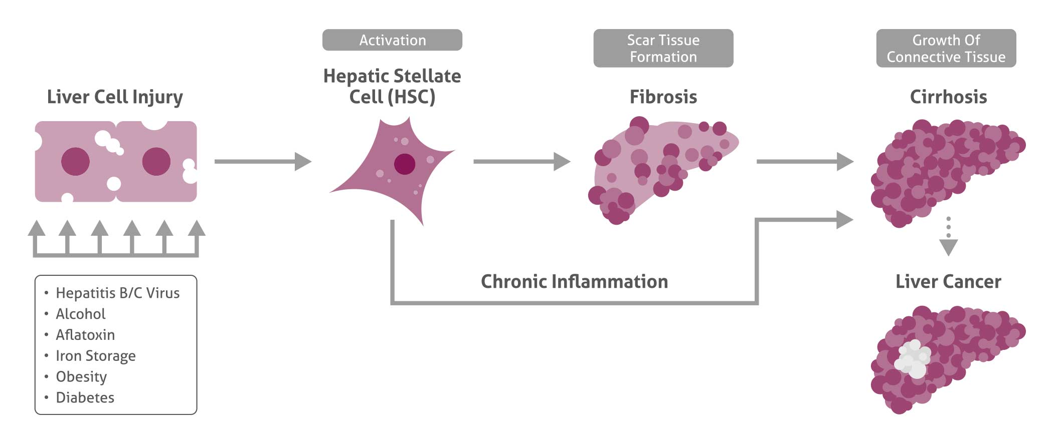
The Many Targets Of The Complex Liver
The liver is the body’s largest glandular organ. This complex organ performs multiple critical functions important for survival (see table 1). The development of all liver diseases and, most importantly, of liver cancer must be understood as a multistep process. Alterations in different signaling pathways or genetic and epigenetic alterations of regulatory genes that lead to uncontrolled in/activation of oncogenes are involved in the development of liver cancer and the exact interactions remain elusive up to this date.3, 4
The mechanism of Wnt signaling has been described in numerous publications. Alterations of the Wnt pathway and its components, e.g., ß-catenin (51067-2- AP) have been proven to be crucial in hepatocarcinogenesis.5, 6 The multifunctional protein ß-catenin is a signaling intermediate that under normal conditions is located in the cytoplasma and continuously degrades. The detection of nuclear ß-catenin by IHC is generally considered to show ß-catenin transcriptional activity. Molecular analysis of human liver and liver-related diseases has also shown in multiple studies the importance of transforming growth factor beta-dependent signaling (TGF-ß) (18978-1-AP)7 via different Smads. TGF-ß is considered to be a central regulator in liver disease, contributing to all stages of disease progression. For instance, Smad7 (25840-1-AP) is known to inhibit TGF-ß signaling via negative feedback loops and Smad3 (25494-1-AP) plays an important role in regulating transcriptional responses. No less important is the Hepatocyte Growth Factor signaling pathway in normal liver, hepatic regeneration, or during tumorgenesis.
| SMAD7 | |
 |
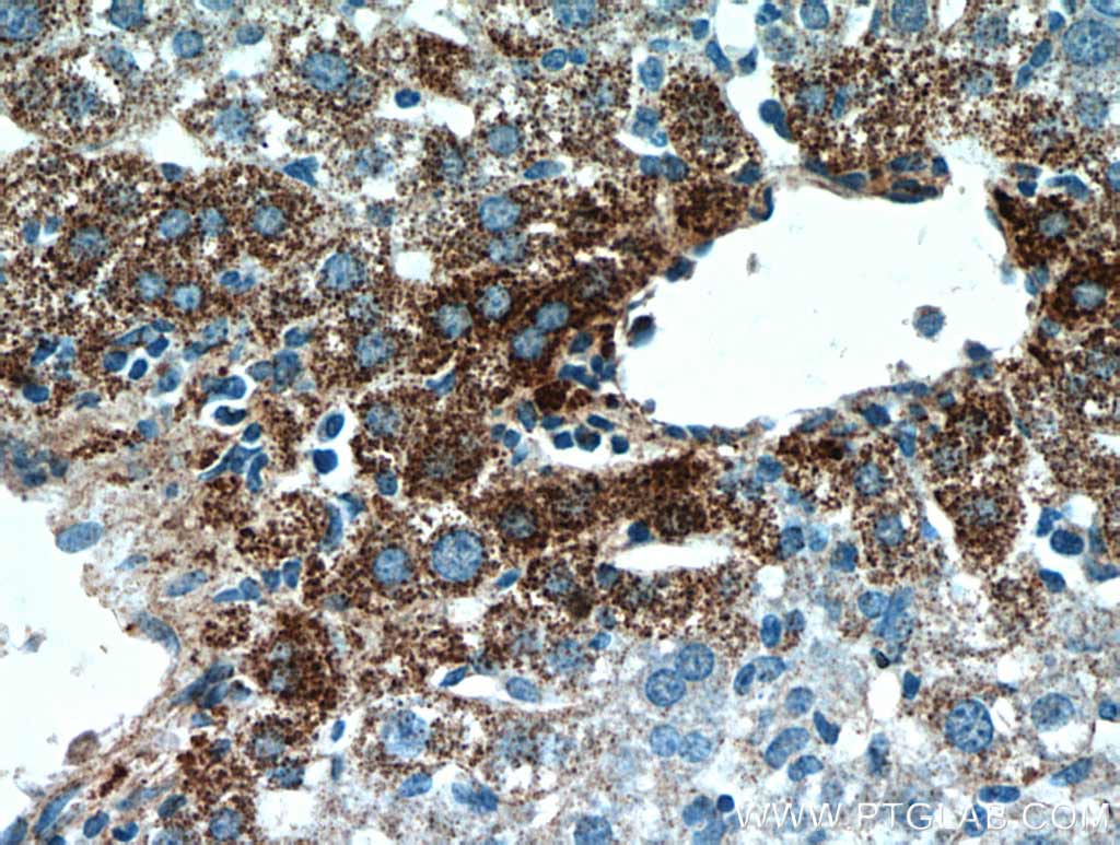
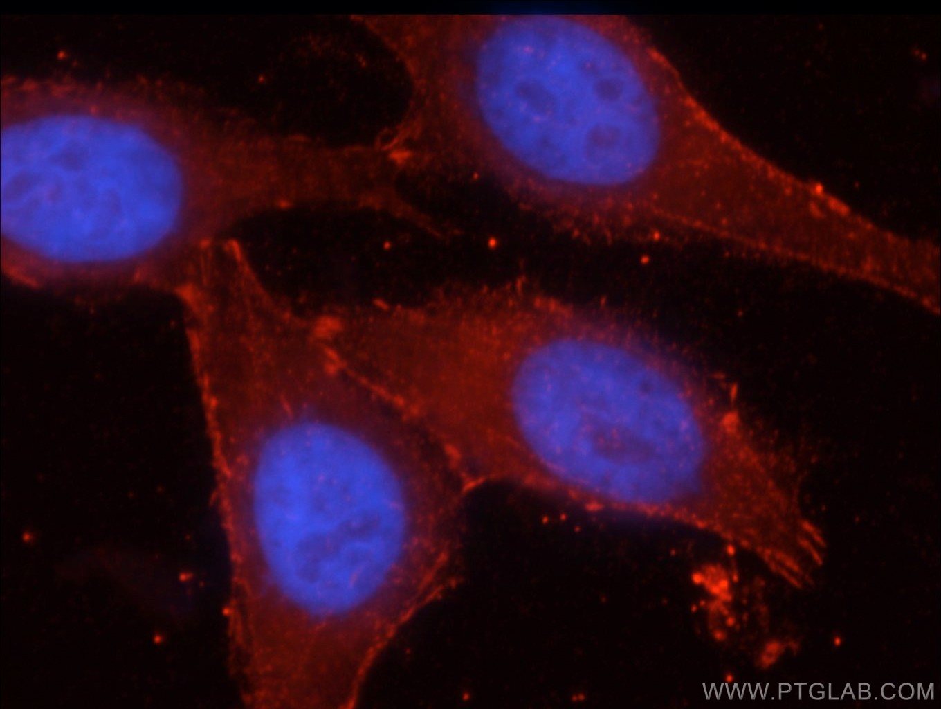 |
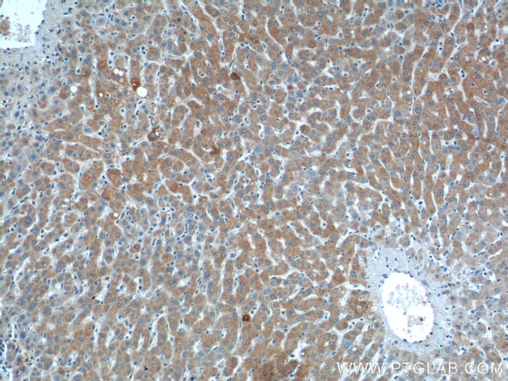
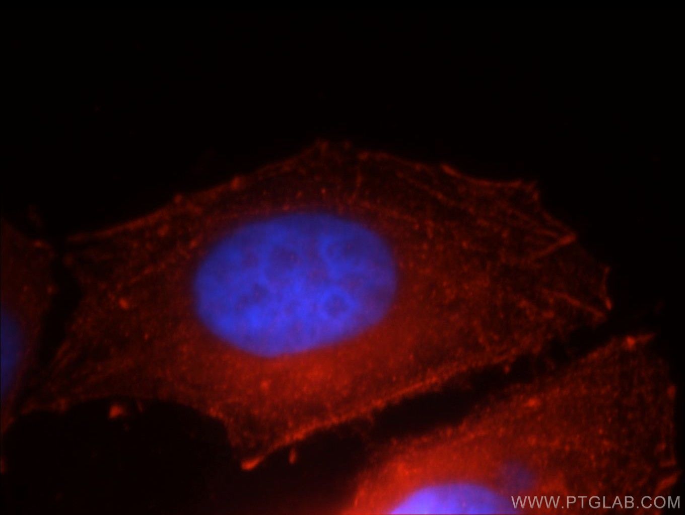
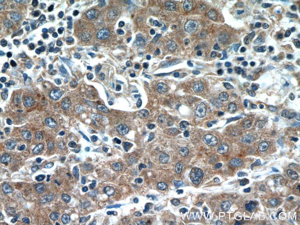
| IHC staining of paraffin-embedded human liver tissue using Smad7 (25840-1-AP)antibody at a dilution of 1:200 (10x objective). | Immunofluorescent analysis of HepG2 cells using beta-catenin (51067-2-AP) antibody at a dilution of 1:50. |
Table 1
| Snapshot Liver Functions |
| Removes harmful substances |
| Stores and digests food |
| Converts food into small substances needed by the body. |
| Synthesizes signaling molecules and plasma proteins. |
Liver Disease infographic: Download here |
Related Products
| Antibody Name | Catalog number | Applications |
| Beta-Catenin | 51067-2-AP | ELISA, IF, IHC, IP, WB |
| Hepatocytes Growth Factor (HGF) | 10390-1-AP | ELISA, IF, IP, WB |
| TGF-ß | 18978-1-AP | ELISA, IHC, WB |
| Smad3 | 25494-1-AP | ELISA, IF, IHC, WB |
| Smad7 | 25840-1-AP | ELISA, WB |
Activation Of Hepatic Stellate Cells
The activation of hepatic stellate cells (HSCs) is considered to be one of the dominant mechanism of fibrogenesis. While in normal liver HSCs are in a quiescent state and serve as the main storage for vitamin A, they become the main matrix-producing cells in the process of liver fibrosis.
The appearance of alpha smooth muscle actin (α-SMA) (14395-1-AP) and desmin (60226-1-Ig, 16520-1-AP) shows the activation of HSCs and are commonly used in immunostaining to identify the stage of HSCs.8, 9 Suitable for double staining is synaptophysin that is instead expressed on the surface of quiescent and activated HSCs.10
Multiple cytokines are involved in the regulation of HSC activation and as the activation of HSC triggers fibrosis, many anti-fibrotic therapies focus on the elucidation of the mechanism driving HSC activation.
Related Products
| Antibody Name | Catalog number | Applications |
| Rabbit Anti Smooth Muscle Actin | 55135-1-AP | ELISA, IHC, WB |
| ACTA2 | 23081-1-AP | ELISA, IHC, IP, WB |
| Desmin | 60226-1-lg | ELISA, IHC, WB |
| Desmin | 16520-1-AP | ELISA, IF, IHC, IP, WB |
| Synaptophysin | 17785-1-AP | ELISA, IF, IHC, IP, WB |
Fibrosis
Liver fibrosis is the result of chronic liver damage, a wound-healing response of the liver to repeated injury. If the hepatic injury persists, liver regeneration fails and apoptotic hepatocytes are substituted by abundant ECM proteins. Excessive accumulation of extracellular matrix (ECM) proteins is therefore the hallmark of liver fibrosis.
For instance, in this stage, the liver contains approximately 6 times more ECM protein-like collagens (Collagen III, 13548-1-AP). During the progression of liver fibrosis, these collagen bands start to bridge and the stage of liver cirrhosis is reached (see next chapter). Activated hepatic stellate cells, portal fibroblasts, or myofibroblasts are known to be the major collagen-producing cells in fibrotic liver (overview of hepatic cells shown in table 2).
Connective Tissue Growth Factor (CTGF, 23936-1-AP) plays a central role in tissue remodeling and it is reported to be a major target promoting epithelial-mesenchymal transition (EMT) during liver fibrosis. CTGF also affects proliferation, differentiation, and ECM synthesis.11, 12, 13 CTGF can act alone or can induce the expression of a variety of cytokines such as Vascular Endothelial Growth Factor (VEGF, 19003-1- AP),14 which again induces more expression of CTGF. VEGF, besides increasing the release of fibrosis-enhancing molecules, has diverse fibrogenic effects; angiogenic function and promoting inflammation.
Liver fibrosis represents the common pathway of chronic liver disease progressing into liver cirrhosis. Continued elucidation of liver fibrosis and its targets will help to provide a complete understanding of the fibrosis mechanism.
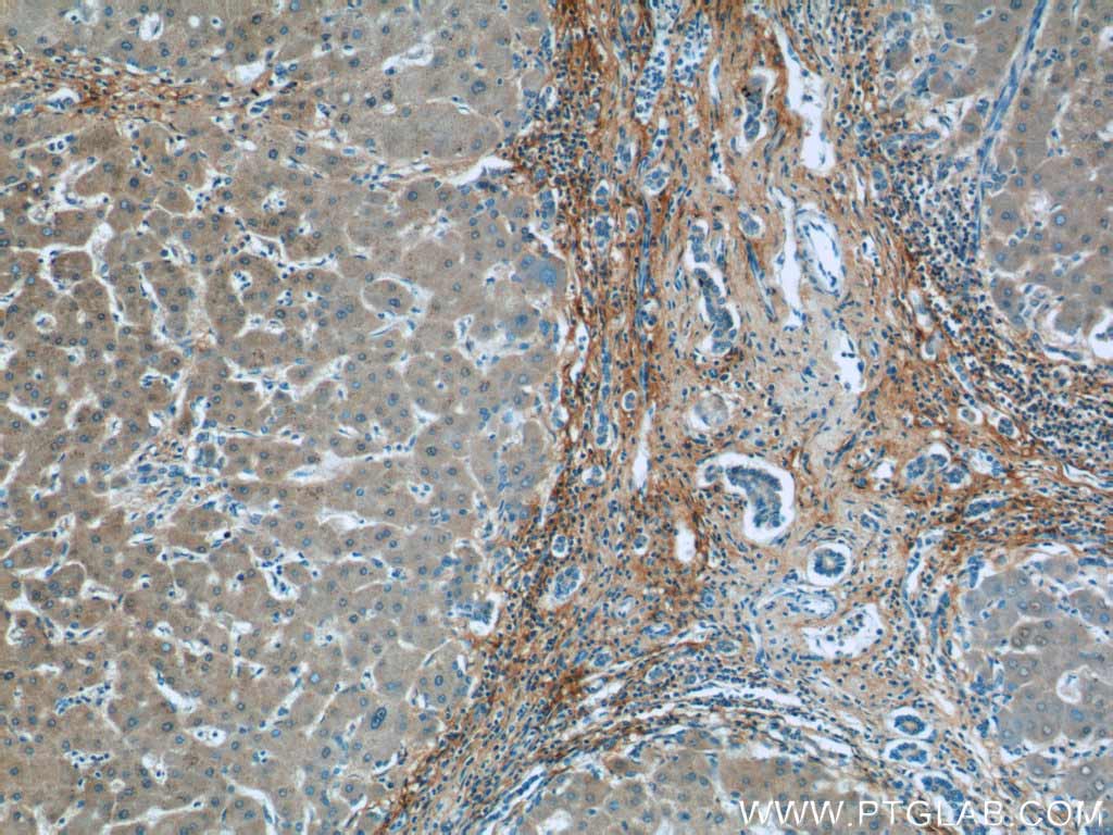
IHC staining of paraffin-embedded human hepatocirrhosis tissue using Collagen III (13548-1-AP) antibody at a dilution of 1:100 (10x objective).IHC staining of paraffin-embedded mouse liver using Collagen III (13548-1-AP) antibody at a dilution of 1:50 (40x objective)
 |
 |
| IHC staining of paraffin-embedded human hepatocirrhosis tissue using Collagen III (13548-1-AP) antibody at a dilution of 1:100 (10x objective) | IHC staining of paraffin-embedded mouse liver using Collagen III (13548-1-AP) antibody at a dilution of 1:50 (40x objective) |
Table 2
| The Liver Comprises Different Cell Types |
| Hepatocytes |
| Hepatic Stellate Cells |
| Progenitor Cells |
| Cholangiocytes |
| Kupffer Cells |
| Endothelial Cells |
Cirrhosis
Advanced liver fibrosis results in cirrhosis. Cirrhosis is characterized by architectural disruption, altered hepatocyte regeneration, and vascular changes. Cirrhosis is also associated with an increased risk of liver failure, portal hypertension, and often requires liver transplantation.15, 16 Because cirrhosis can lead to life-threatening complications, its accurate assessment is important. The high contribution of inflammatory signaling driving chronic liver disease remains incompletely understood, to name just one here, Lymphotoxin-ß receptor (LTßR) (20331-1-AP) mediated signaling. It has been reported to be overexpressed in chronic liver damage in multiple hepatic cell types when fibrosis or cirrhosis is present17 and activation of the LT-ß receptor induces production of chemokines and adhesion molecules promoting inflammation.18
In addition, hepatic angiogenesis and capillarization of the sinusoids are the main features during the progression of cirrhosis. Expression of CD34 (14486-1-AP) positive endothelial cells plays an important role in understanding the process of angiogenesis in cirrhosis.19 Sinusoidal capillarization goes along with formation of tight junctions between endothelial cells and formation of new vessels. CD31 (11265-1-AP) is a specific and sensitive marker to detect capillary units.19 Understanding the processes of cirrhosis might help in the design of efficient therapy for this liver disorder before it develops further into cancer. Go to www.ptglab.com and pick your marker of interest.
Related Products
| Antibody Name | Catalog Number | Applications |
| LTBR | 20331-1-AP | ELISA, FC, IHC, WB |
| CD34 | 60180-1-Ig | ELISA, IHC, WB |
| CD31 | 11265-1-AP | ELISA, IHC, WB |
Liver Cancer
Hepatocellular carcinoma (HCC) is a leading cause of death, being the fifth most common cancer in the world. Since it is rapidly progressing and therapies are limited, diagnosis and intervention at an early stage are essential.
Commonly used biomarkers are, for instance, α–fetoprotein (AFP) (14550-1-AP), albumin (16475-1-AP), arginase-1 (16001-1-AP, 66129- 1-Ig), vimentin (10366-1-AP), (17513-1-AP), Carbonic anhydrase 9 (11071-1-AP, 66243- 1-Ig), glutamine synthetase (11037-2-AP), or E-cadherin (20648-1-AP). However, due to the complexity of the disease, new factors are constantly reported in order to better understand the pathogenesis of the disease.
A new marker has been reported with the Golgi protein-73 (GP73) that is normally expressed in epithelial cells of many human tissues. It is essential for human survival, and has multiple cell functions in the liver. GP73 expression is upregulated in serum samples from patients with liver disease, with expression being highest in HCC.20
Thyroid transcription factor (TTF)-1 (66034-1-Ig) is also a very useful tumor marker discovered during the past few years that might help in distinguishing primary carcinoma from metastatic liver carcinoma.21 High levels of S100A6 (10245-1-AP) and FUCA1 (16420-1-AP) have been associated with poor outcome, promoting cell proliferation and migration in human hepatocellular carcinoma.22, 23 Glypican 3 (GPC3) (25175-1-AP), for instance, has recently been reported to be a novel marker of hepatocellular carcinoma (HCC), suggesting tumor growth inhibition dependent on GPC3 and its potential as a therapeutic target for immunotherapy.24
During the last several years, major progress has been achieved with regard to early diagnosis and increasing knowledge of molecular hepatocarcinogenesis. These achievements help to provide new opportunities for targeted therapy.
 |
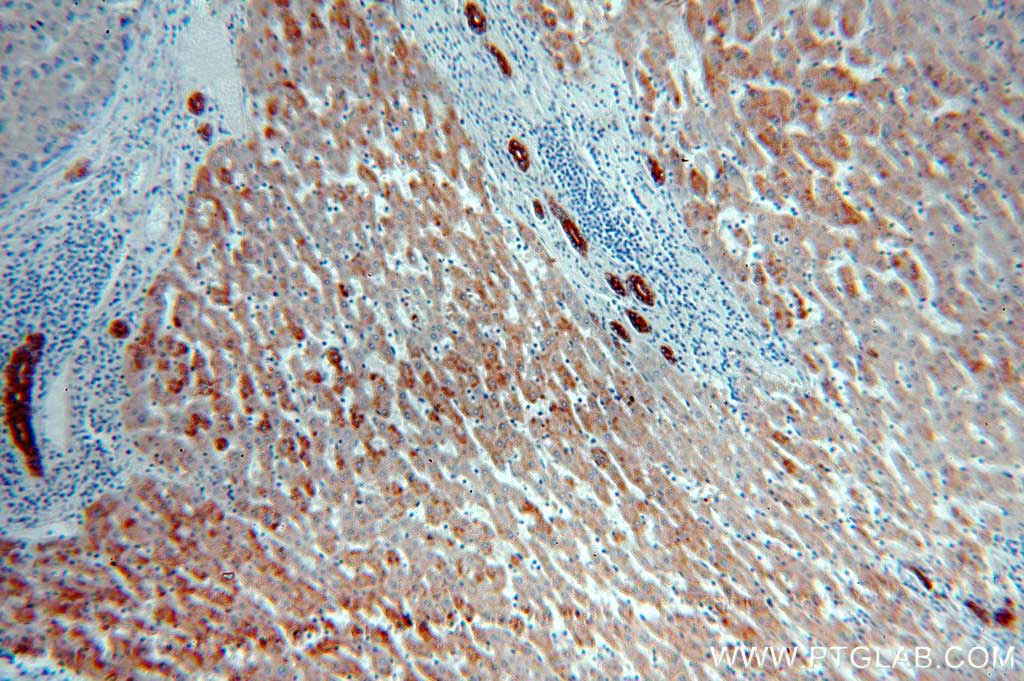 |
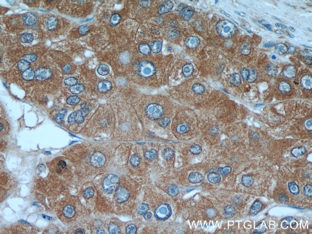
| IHC staining of paraffin-embedded human liver cancer using α-Fetoprotein (AFP, 14550-1-AP) at a dilution of 1:50 (40x objective). | IHC staining of paraffin-embedded human liver using Cytokeratin specific (CK7, 17513-1-AP) antibody at a dilution of 1:50 (10x objective). |
 |
 |
| Immunofluorescent analysis of HepG2 cells using E-Cadherin (20648-1-AP) antibody at a dilution of 1:25 and Rhodamine-labelled goat anti-rabbit IgG (red), DAPI (blue). | IHC staining of paraffin-embedded human liver cancer tissue using Glypican3 (GPC3, 25175-1-AP) antibody at a dilution of 1:50 (40x objective). |
Related Products
| Antibody Name | Catalog number | Applications |
| α-Fetoprotein (AFP) | 14550-1-AP | ELISA, FC, IF, IHC, IP, WB |
| Albumin | 16475-1-AP | ELISA, FC, IF, IHC, WB |
| Arginase-1 | 16001-1-AP | ELISA, FC, IF, IHC, IP, WB |
| Arginase-1 | 66129-1-lg | ELISA, IHC, WB |
| CA9 | 11071-1-AP | ELISA, FC, IF, IHC, WB |
| CA9 | 66243-1-lg | ELISA, IF, IHC, WB |
| Cytokeratin 7 | 17513-1-AP | ELISA, IF, IHC, IP, WB |
| E-Cadherin | 20648-1-lg | ELISA, FC, IF, IHC, WB |
| FUCA1 | 16420-1-AP | ELISA, IHC |
| Glutamine Synthetase | 11037-2-AP | ELISA, FC, IF, IHC, IP, WB |
| Glypican 3 (GPC3) | 25175-1-AP | ELISA, IF, IHC, WB |
| GP73 | 15126-1-AP | ELISA, FC, IF, IHC, IP, WB |
| HSP70 | 10995-1-AP | ELISA, FC, IF, IHC, IP, WB |
| S100A6 | 10245-1-AP | ELISA, FC, IF, IHC, IP, WB |
| TTF-1 | 66034-1-lg | ELISA, IHC, WB |
| Vimentin | 10366-1-AP | ELISA, FC, IF, IHC, WB |
Download this product focus as a PDF |
Click here for the full list of references
Loading control antibodies
| GAPDH Antibody |  |
| Catalog no.: 60004-1-Ig | |
|
GAPDH is commonly used as a protein loading control in western blot due to its consistently high expression in most cell types. This enzyme participates in several cellular events such as glycolysis, DNA repair, and apoptosis. Proteintech monoclonal GAPDH antibodies are raised against a whole-protein antigen of human origin and have over 4,960 citations. |
| Beta Actin Antibody (KD/KO validated) |  |
| Catalog no.: 66009-1-Ig | |
|
Beta-actin is usually used as a loading control due to its broad and consistent expression across all eukaryotic cell types and the fact that expression levels of this protein are not affected by most experimental treatments. 66009-1-Ig has been cited in over 2,460 publications and has wide species reactivity. |


