Proteintech修饰性抗体优势
-

高特异性从源头开始严格抗原设计以及负向筛选确保高特异性
-
匹配真实应用场景内源性免疫学实验验证及刺激样本验证确保应用数据真实
修饰性抗体检测数据
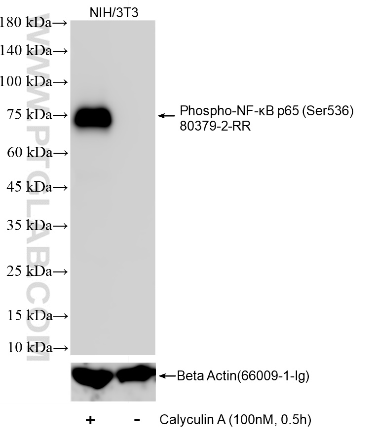
Non-treated and Calyculin A treated NIH/3T3 cells were subjected to SDS PAGE followed by western blot with 80379-2-RR (Phospho-NF-κB p65 (Ser536) antibody) at dilution of 1:5000 incubated at room temperature for 1.5 hours. The membrane was stripped and re-blotted with Beta Actin (66009-1-Ig) antibody as a loading control.
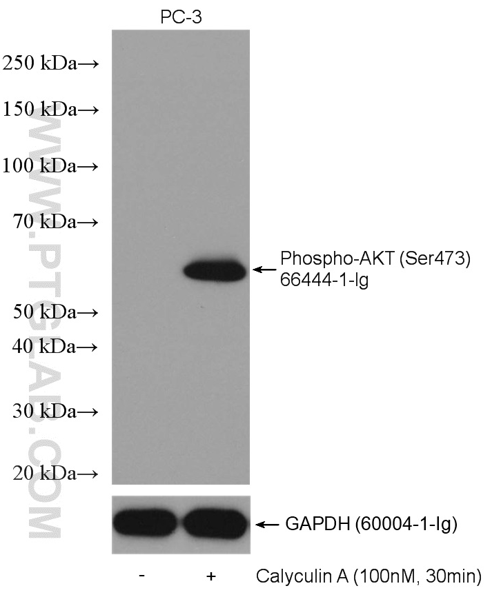
Non-treated PC-3 and Calyculin A treated PC-3 cells were subjected to SDS PAGE followed by western blot with 66444-1-Ig (Phospho-AKT (Ser473) antibody) at dilution of 1:5000 incubated at room temperature for 1.5 hours. The membrane was stripped and re-blotted with GAPDH antibody as loading control.
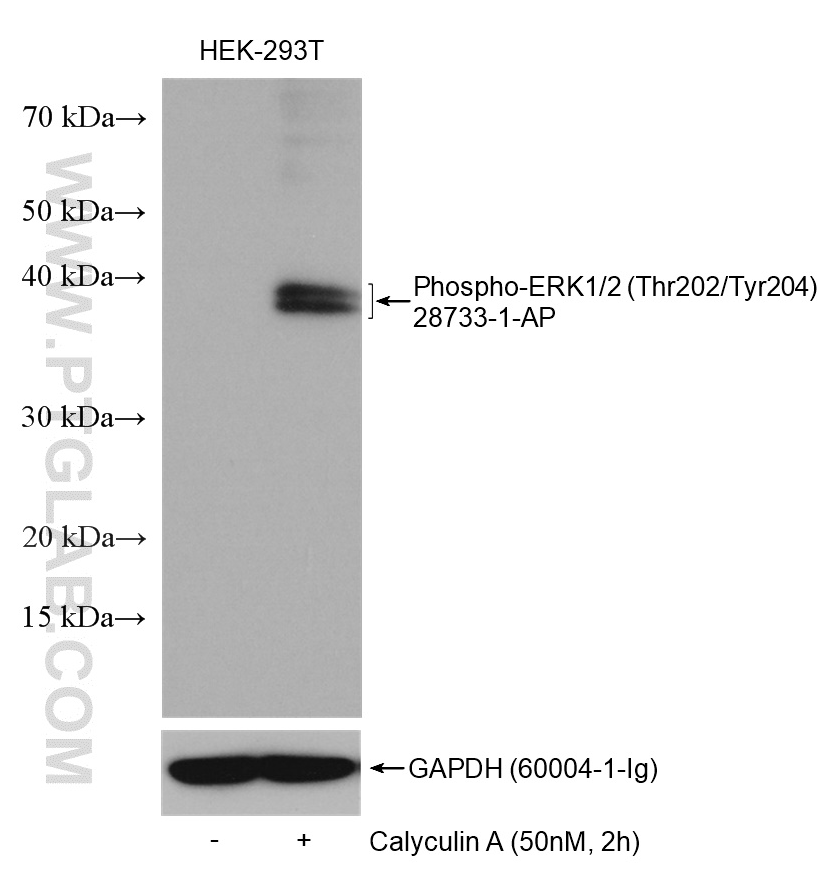
Non-treated HEK-293T and Calyculin A treated HEK-293T cells were subjected to SDS PAGE followed by western blot with 28733-1-AP (ERK1/2-phospho-Thr202/Tyr204) at dilution of 1:3000 incubated at 4℃ overnight. The membrane was stripped and re-blotted with GAPDH antibody as loading control.
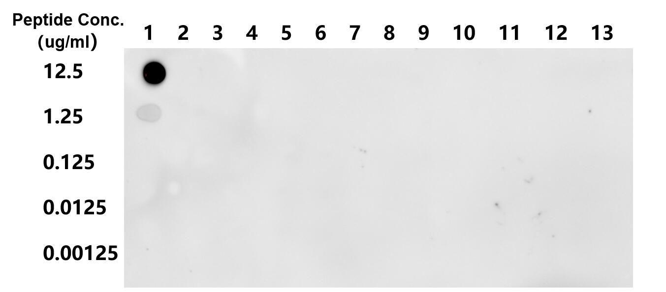
Dot blot analysis was used to confirm the specificity of Acetyl-Histone H3 (Lys27) antibody. Acetylated peptides were spotted onto NC and probed with antibody at 1 µg/ml.The amount of peptide (ug/mL) spotted is indicated next to each row. Column 1: H3K27ac. Column 2: Unmodified H3K27. Column 3: H3K9ac. Column 4: H3K14ac. Column 5: H3K18ac. Column 6: H3K23ac. Column 7: H3K36ac. Column 8: H4K5ac. Column 9: H4K8ac. Column 10: H4K12ac. Column 11: H4K16ac. Column 12: H2AK5ac. Column 13: Blank(PBS).
Chromatin was prepared from HeLa cells. Cells were fixed with formaldehyde for 10 minutes. The ChIP was performed with 15 µg of cross-linked chromatin, 5 µg of Acetyl-Histone H3 (Lys4) (84831-1-RR) or 5 ug of Normal Rabbit IgG (98136-1-RR), and 20 µl of Protein A Magarose Beads. The immunoprecipitated DNA was quantified by real-time PCR.
Dot blot analysis was used to confirm the specificity of 84549-1-RR Acetyl-Histone H2B (Lys20) antibody. Peptides were spotted onto NC and probed with antibody at 1 µg/ml. The amount of peptide (ug/mL) spotted is indicated next to each row.
Chromatin was prepared from HeLa cells. Cells were fixed with formaldehyde for 10 minutes. The ChIP was performed with 15 µg of cross-linked chromatin, 5 µg of Mono-Methyl-Histone H3 (Lys36) (82825-2-RR) or 5 ug of Normal Rabbit IgG (98136-1-RR), and 20 µl of Protein A Magarose Beads. The immunoprecipitated DNA was quantified by real-time PCR.
Di-Methyl-Histone H3(Lys36) antibody (29202-1-AP) tested by ELISA. HIST1H3A(31-43aa), HIST1H3AMe1(31-43aa), HIST1H3AMe2(31-43aa), HIST1H3AMe3(31-43aa) were coated onto microtiter plates at 0.15 µg/well and then incubated with a dilution series of HIST1H3AMe2 antibody (29202-1-AP). Bound antibodies were detected with HRP conjugated anti-rabbit IgG followed by incubation with HRP substrate and then measuring the resulting absorbance at 450 nm.

Chromatin was prepared from HeLa cells. Cells were fixed with formaldehyde for 10 minutes. The ChIP was performed with 10 µg of cross-linked chromatin, 3 µg of Di/Tri-Methyl-Histone H3 (Lys4) (84908-2-RR) or 3 ug of Normal Rabbit IgG (98136-1-RR), and 20 µl of Protein A Magarose Beads. The immunoprecipitated DNA was quantified by real-time PCR.
表观遗传学研究相关修饰抗体
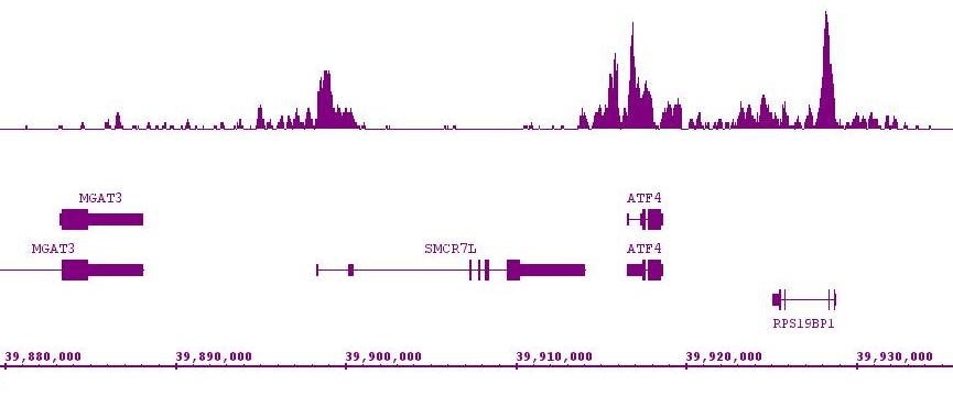
Histone H3K4me3 antibody (mAb) tested by ChIP-Seq. ChIP was performed using the ChIP-IT High Sensitivity Kit (Cat. No. 53040) with 15 ug of chromatin from a human medulloblastoma cell line and 4 ug of antibody. ChIP DNA was sequenced on the Illumina HiSeq and 6 million sequence tags were mapped to identify Histone H3K4me3 binding sites. The image shows binding across a region of chromosome 7. You can view the complete data set in the UCSC Genome Browser, starting at this specific location, here
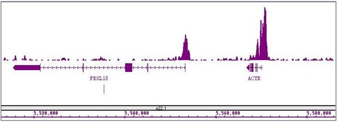
Histone H3K9ac antibody (mAb) (Clone 2G1F9) tested by ChIP-Seq. ChIP was performed using the ChIP-IT High Sensitivity Kit (Cat. No. 53040) with 15 ug of chromatin from a human medulloblastoma cell line and 4 ug of antibody. ChIP DNA was sequenced on the Illumina HiSeq and 6 million sequence tags were mapped to identify Histone H3K9ac binding sites. The image shows binding across a region of chromosome 22. You can view the complete data set in the UCSC Genome Browser, starting at this specific location, here.


