验证数据展示


经过测试的应用
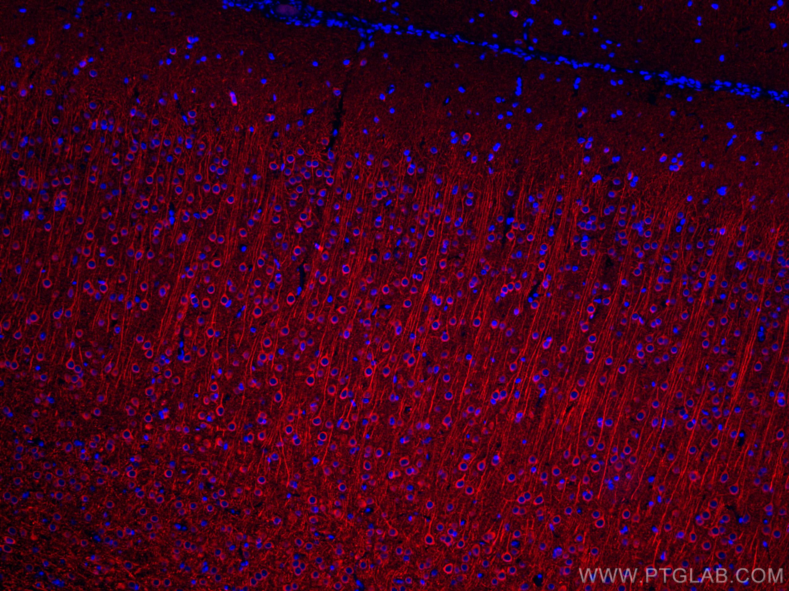
| Positive IF-P detected in | rat brain tissue |
| Positive FC (Intra) detected in | Neuro-2a cells |
推荐稀释比
| 应用 | 推荐稀释比 |
|---|---|
| Immunofluorescence (IF)-P | IF-P : 1:50-1:500 |
| Flow Cytometry (FC) (INTRA) | FC (INTRA) : 0.40 ug per 10^6 cells in a 100 µl suspension |
| It is recommended that this reagent should be titrated in each testing system to obtain optimal results. | |
| Sample-dependent, Check data in validation data gallery. | |
发表文章中的应用
| IF | See 2 publications below |
产品信息
CL594-17490 targets MAP2 in IF-P, FC (Intra) applications and shows reactivity with human, mouse, rat samples.
| 经测试应用 | IF-P, FC (Intra) Application Description |
| 文献引用应用 | IF |
| 经测试反应性 | human, mouse, rat |
| 文献引用反应性 | human, mouse, rat |
| 免疫原 |
CatNo: Ag11580 Product name: Recombinant human MAP2 protein Source: e coli.-derived, PGEX-4T Tag: GST Domain: 213-559 aa of BC038857 Sequence: TSAGSTDRLPYSKSGNKDGVTKSPEKRSSLPRPSSILPPRRGVSGDRDENSFSLNSSISSSARRTTRSEPIRRAGKSGTSTPTTPGSTAITPGTPPSYSSRTPGTPGTPSYPRTPHTPGTPKSAILVPSEKKVAIIRTPPKSPATPKQLRLINQPLPDLKNVKSKIGSTDNIKYQPKGGQVRILNKKIDFSKVQSRCGSKDNIKHSAGGGNVQIVTKKIDLSHVTSKCGSLKNIRHRPGGGRVKIESVKLDFKEKAQAKVGSLDNAHHVPGGGNVKIDSQKLNFREHAKARVDHGAEIITQSPGRSSVASPRRLSNVSSSGSINLLESPQLATLAEDVTAALAKQGL 种属同源性预测 |
| 宿主/亚型 | Rabbit / IgG |
| 抗体类别 | Polyclonal |
| 产品类型 | Antibody |
| 全称 | microtubule-associated protein 2 |
| 别名 | MAP 2, MAP-2, MAP2A, MAP2B, MAP2C |
| 计算分子量 | 200 kDa |
| GenBank蛋白编号 | BC038857 |
| 基因名称 | MAP2 |
| Gene ID (NCBI) | 4133 |
| RRID | AB_2919842 |
| 偶联类型 | CoraLite®594 Fluorescent Dye |
| 最大激发/发射波长 | 588 nm / 604 nm |
| 激发激光 | Yellow-Green Laser (561 nm) |
| 形式 | Liquid |
| 纯化方式 | Antigen affinity purification |
| UNIPROT ID | P11137 |
| 储存缓冲液 | PBS with 50% glycerol, 0.05% Proclin300, 0.5% BSA, pH 7.3. |
| 储存条件 | Store at -20°C. Avoid exposure to light. Stable for one year after shipment. Aliquoting is unnecessary for -20oC storage. |
背景介绍
Microtubule-associated protein 2 (MAP2) is a tubulin binding protein regulating the spacing and stability of microtubules and contributing to elongation of dendrites.
What is the molecular weight of MAP2? Is MAP2 post-translationally modified?
MAP2 has multiple isoforms that arise from alternative splicing (PMID: 3121794, 7854050, and 10383434). They are classified into two groups - MAP2A and MAP2B, which are known as high molecular weight (HMW) isoforms, run as ~280 kDa species, while low molecular weight (LMW) isoforms MAP2C and MAP2D are around ~70 kDa. MAP2 proteins are heavily phosphorylated, which contributes to a large discrepancy between their predicted and observed molecular weight in SDS-PAGE (220 vs 280 kDa for HMW forms).
What is the tissue expression pattern of MAP2? What is the subcellular localization of MAP2?
MAP2 isoforms differ in their tissue and developmental expression pattern (PMID: 2469170 and 3898077). In the brain, MAP2B is widely expressed during and post development, MAP2A is expressed postnatally, while MAP2C is present only in the early development except of present in photosensitive cells of the adult retina and in the olfactory system. MAP proteins are highly expressed in the CNS found in cell bodies and dendrites of neurons, in dorsal root ganglion, reactive glia, and in the testis (PMID: 9588626). In neurons, MAP2 proteins are found in the cell body and dendrites, where they associate with microtubules, while they can also be present in the nuclei of testicular cells.
Can MAP2 be used as a neuronal marker?
MAP2 proteins are abundantly expressed in neurons. MAP2 is frequently used as a dendritic marker because it is present in the cell body and dendrites of neurons but absent in axons (PMID: 28413822).
实验方案
| Product Specific Protocols | |
|---|---|
| FC protocol for CL594 MAP2 antibody CL594-17490 | Download protocol |
| IF protocol for CL594 MAP2 antibody CL594-17490 | Download protocol |
| Standard Protocols | |
|---|---|
| Click here to view our Standard Protocols |
发表文章
| Species | Application | Title |
|---|---|---|
Biomolecules Interplay between Energy Supply and Glutamate Toxicity in the Primary Cortical Culture | ||
Acta Pharm Sin B Drofenine as a Kv2.1 inhibitor alleviated AD-like pathology in mice through Aβ/Kv2.1/microglial NLRP3/neuronal Tau axis | ||
Mol Psychiatry Frontotemporal dementia patient-derived iPSC neurons show cell pathological hallmarks and evidence for synaptic dysfunction and DNA damage. |