WB Figures
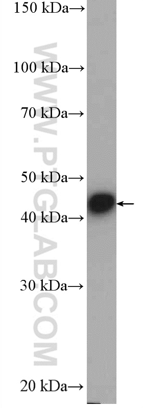
WB analysis of HEK-293 using 10582-1-AP (same clone as 10582-1-PBS)
HEK-293 cells were subjected to SDS PAGE followed by western blot with 10582-1-AP (PELO antibody) at dilution of 1:500 incubated at room temperature for 1.5 hours. This data was developed using the same antibody clone with 10582-1-PBS in a different storage buffer formulation.
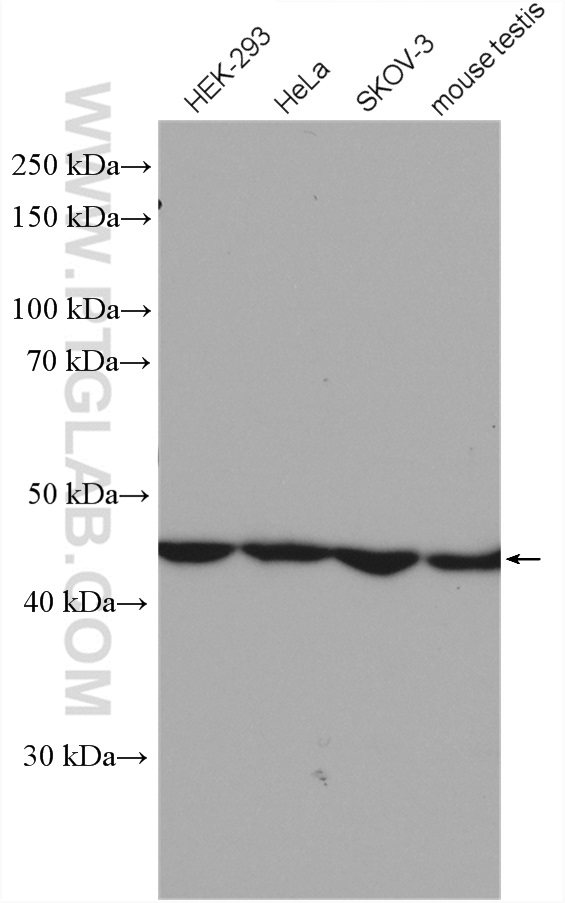
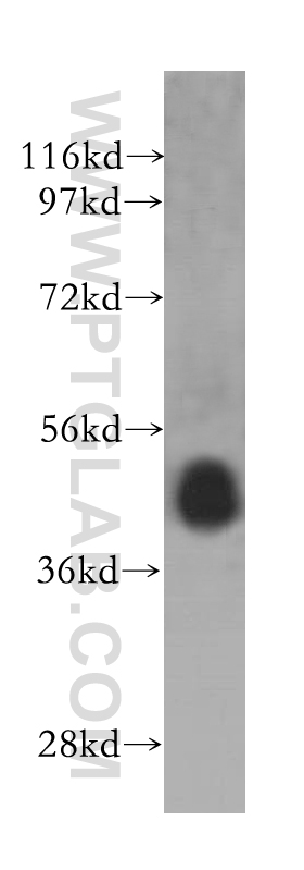
WB analysis using 10582-1-AP (same clone as 10582-1-PBS)
Various cells were subjected to SDS PAGE followed by western blot with 10582-1-AP (PELO antibody) at dilution of 1:3000 incubated at room temperature for 1.5 hours. This data was developed using the same antibody clone with 10582-1-PBS in a different storage buffer formulation.
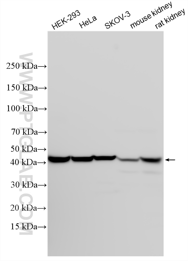
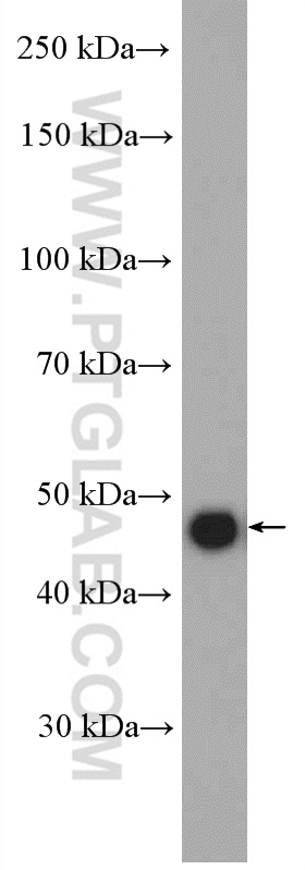
WB analysis using 10582-1-AP (same clone as 10582-1-PBS)
Various lysates were subjected to SDS PAGE followed by western blot with 10582-1-AP (PELO antibody) at dilution of 1:6000 incubated at room temperature for 1.5 hours. This data was developed using the same antibody clone with 10582-1-PBS in a different storage buffer formulation.
WB analysis of HeLa using 10582-1-AP (same clone as 10582-1-PBS)
HeLa cells were subjected to SDS PAGE followed by western blot with 10582-1-AP (PELO antibody) at dilution of 1:1000 incubated at room temperature for 1.5 hours. This data was developed using the same antibody clone with 10582-1-PBS in a different storage buffer formulation.
WB analysis of mouse testis using 10582-1-AP (same clone as 10582-1-PBS)
mouse testis tissue were subjected to SDS PAGE followed by western blot with 10582-1-AP (PELO antibody) at dilution of 1:1000 incubated at room temperature for 1.5 hours. This data was developed using the same antibody clone with 10582-1-PBS in a different storage buffer formulation.
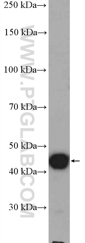
WB analysis of mouse testis using 10582-1-AP (same clone as 10582-1-PBS)
mouse testis tissue were subjected to SDS PAGE followed by western blot with 10582-1-AP (PELO antibody) at dilution of 1:3000 incubated at room temperature for 1.5 hours. This data was developed using the same antibody clone with 10582-1-PBS in a different storage buffer formulation.
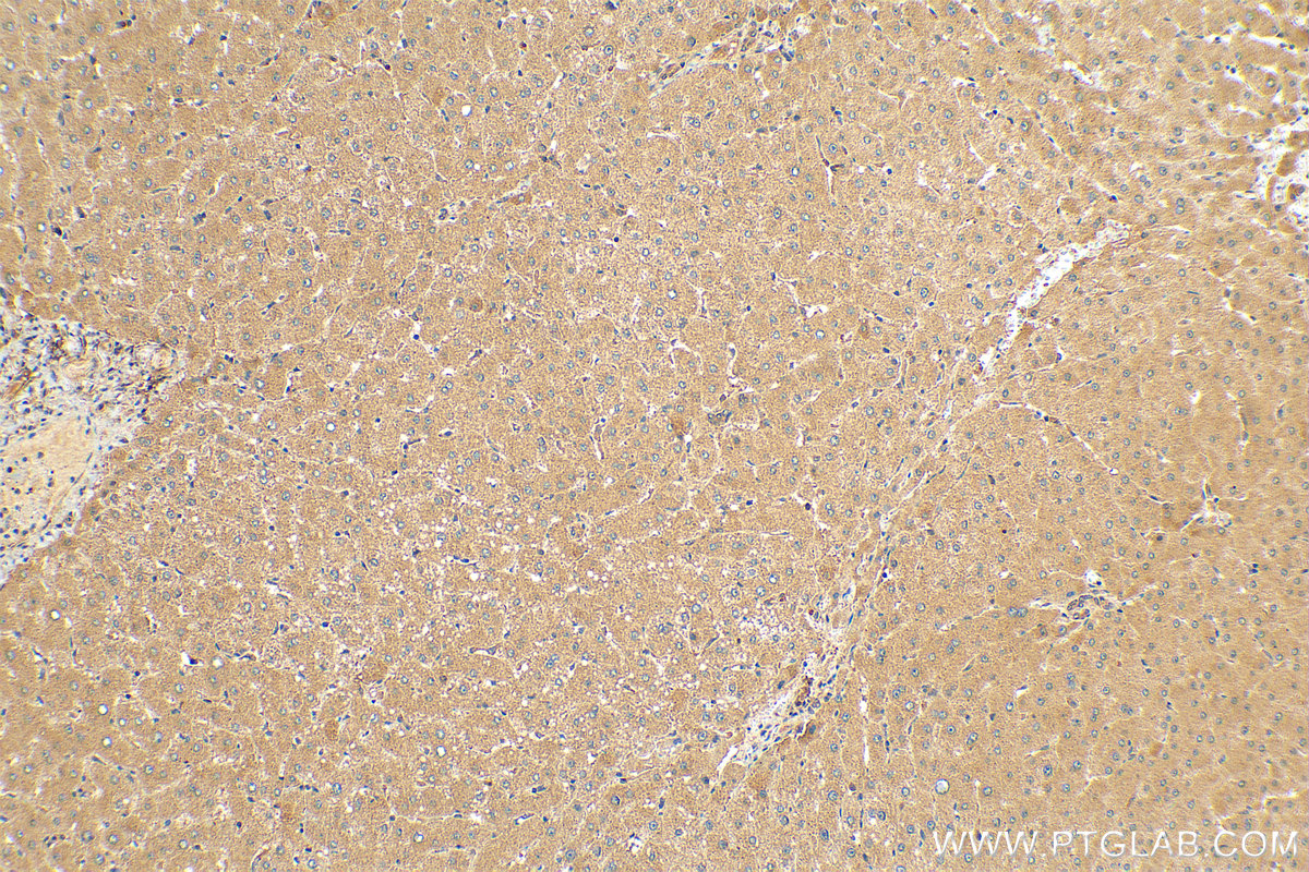
IHC staining of human liver using 10582-1-AP (same clone as 10582-1-PBS)
Immunohistochemical analysis of paraffin-embedded human liver tissue slide using 10582-1-AP (PELO antibody) at dilution of 1:200 (under 10x lens). Heat mediated antigen retrieval with Tris-EDTA buffer (pH 9.0). This data was developed using the same antibody clone with 10582-1-PBS in a different storage buffer formulation.
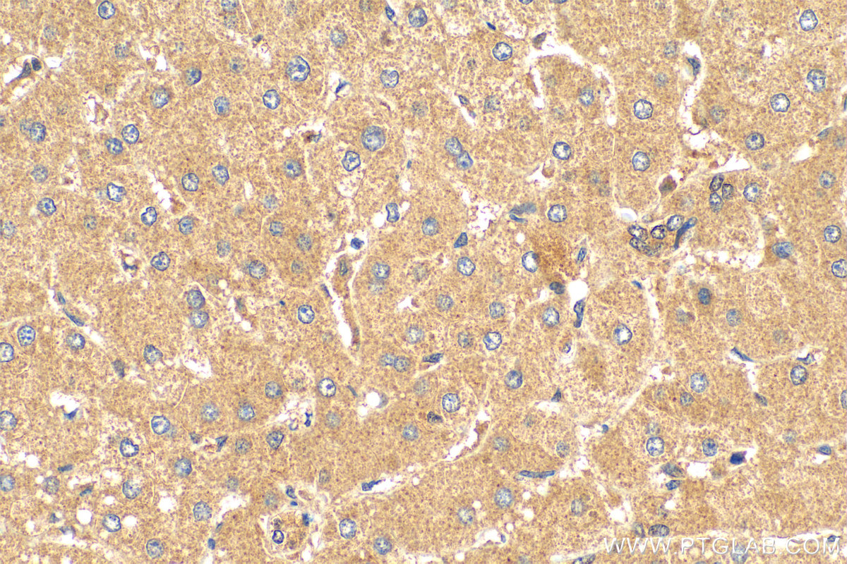
IHC staining of human liver using 10582-1-AP (same clone as 10582-1-PBS)
Immunohistochemical analysis of paraffin-embedded human liver tissue slide using 10582-1-AP (PELO antibody) at dilution of 1:200 (under 40x lens). Heat mediated antigen retrieval with Tris-EDTA buffer (pH 9.0). This data was developed using the same antibody clone with 10582-1-PBS in a different storage buffer formulation.
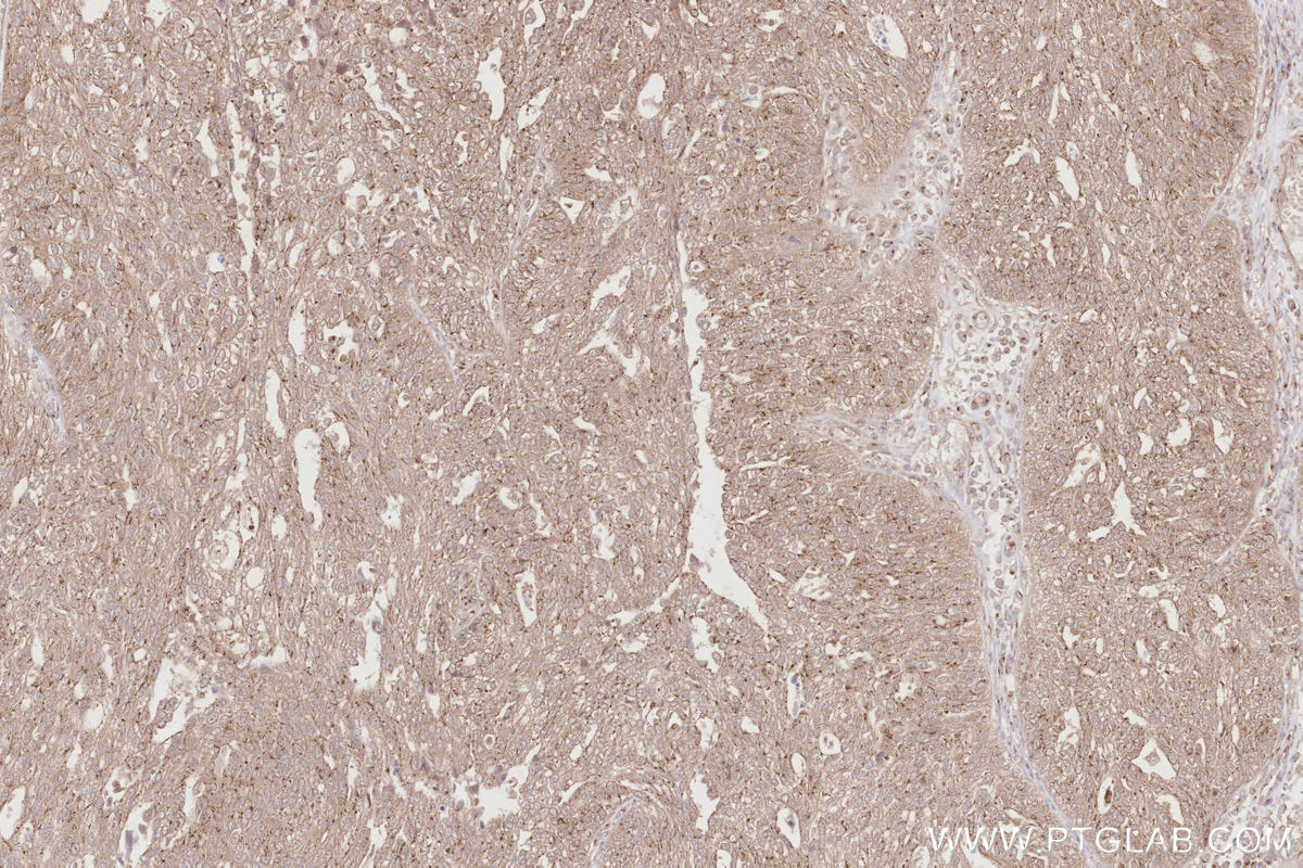
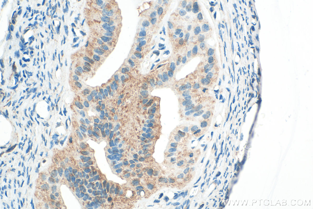
IHC staining of human ovary cancer using 10582-1-AP (same clone as 10582-1-PBS)
Immunohistochemical analysis of paraffin-embedded human ovary cancer tissue slide using 10582-1-AP (PELO antibody) at dilution of 1:500 (under 10x lens). Heat mediated antigen retrieval with Tris-EDTA buffer (pH 9.0). This data was developed using the same antibody clone with 10582-1-PBS in a different storage buffer formulation.
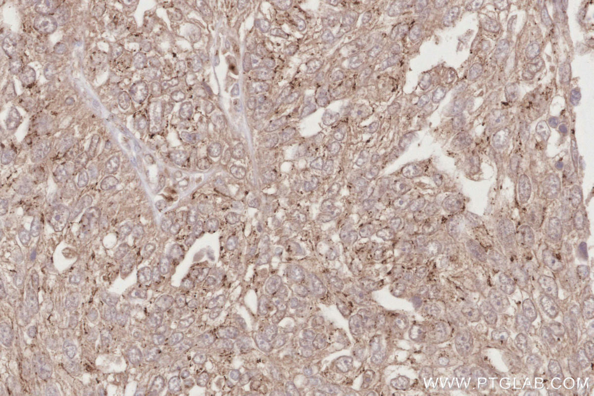
IHC staining of human ovary cancer using 10582-1-AP (same clone as 10582-1-PBS)
Immunohistochemical analysis of paraffin-embedded human ovary cancer tissue slide using 10582-1-AP (PELO antibody) at dilution of 1:500 (under 40x lens). Heat mediated antigen retrieval with Tris-EDTA buffer (pH 9.0). This data was developed using the same antibody clone with 10582-1-PBS in a different storage buffer formulation.
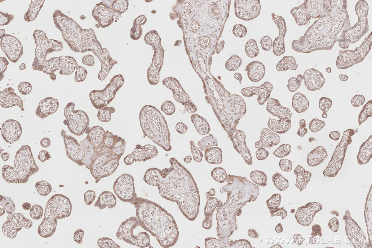
IHC staining of human placenta using 10582-1-AP (same clone as 10582-1-PBS)
Immunohistochemical analysis of paraffin-embedded human placenta tissue slide using 10582-1-AP (PELO antibody) at dilution of 1:500 (under 10x lens). Heat mediated antigen retrieval with Tris-EDTA buffer (pH 9.0). This data was developed using the same antibody clone with 10582-1-PBS in a different storage buffer formulation.
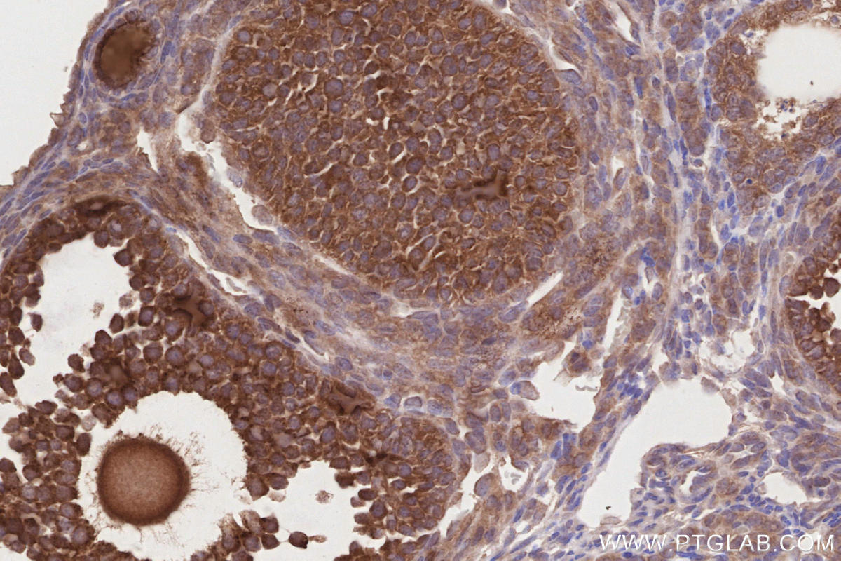
IHC staining of mouse ovary using 10582-1-AP (same clone as 10582-1-PBS)
Immunohistochemical analysis of paraffin-embedded mouse ovary tissue slide using 10582-1-AP (PELO antibody) at dilution of 1:500 (under 40x lens). Heat mediated antigen retrieval with Tris-EDTA buffer (pH 9.0). This data was developed using the same antibody clone with 10582-1-PBS in a different storage buffer formulation.
IHC staining of mouse ovary using 10582-1-AP (same clone as 10582-1-PBS)
Immunohistochemical analysis of paraffin-embedded mouse ovary tissue slide using 10582-1-AP (PELO antibody) at dilution of 1:500 (under 40x lens). Heat mediated antigen retrieval with Tris-EDTA buffer (pH 9.0). This data was developed using the same antibody clone with 10582-1-PBS in a different storage buffer formulation.
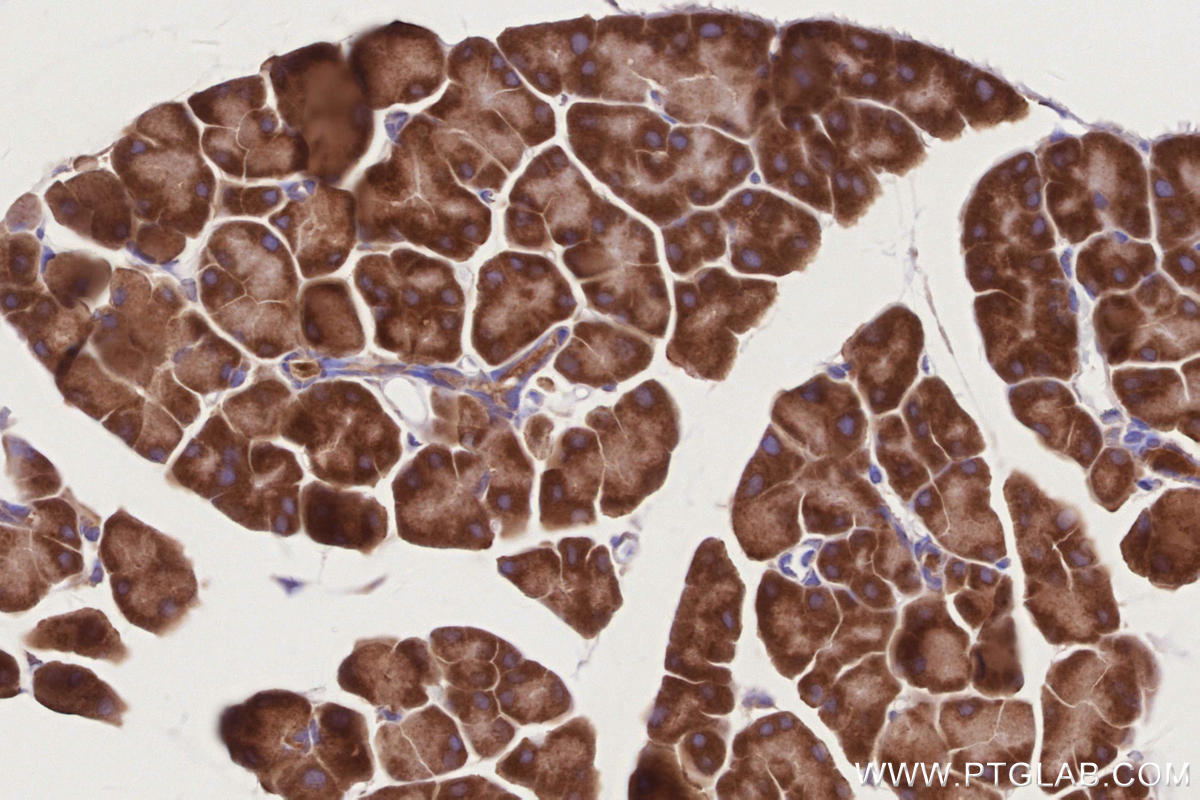
IHC staining of mouse pancreas using 10582-1-AP (same clone as 10582-1-PBS)
Immunohistochemical analysis of paraffin-embedded mouse pancreas tissue slide using 10582-1-AP (PELO antibody) at dilution of 1:500 (under 40x lens). Heat mediated antigen retrieval with Tris-EDTA buffer (pH 9.0). This data was developed using the same antibody clone with 10582-1-PBS in a different storage buffer formulation.
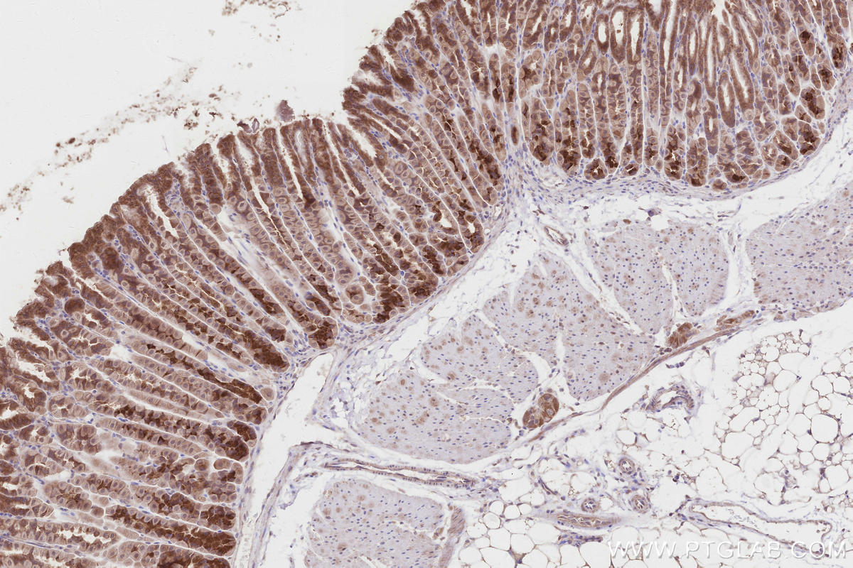
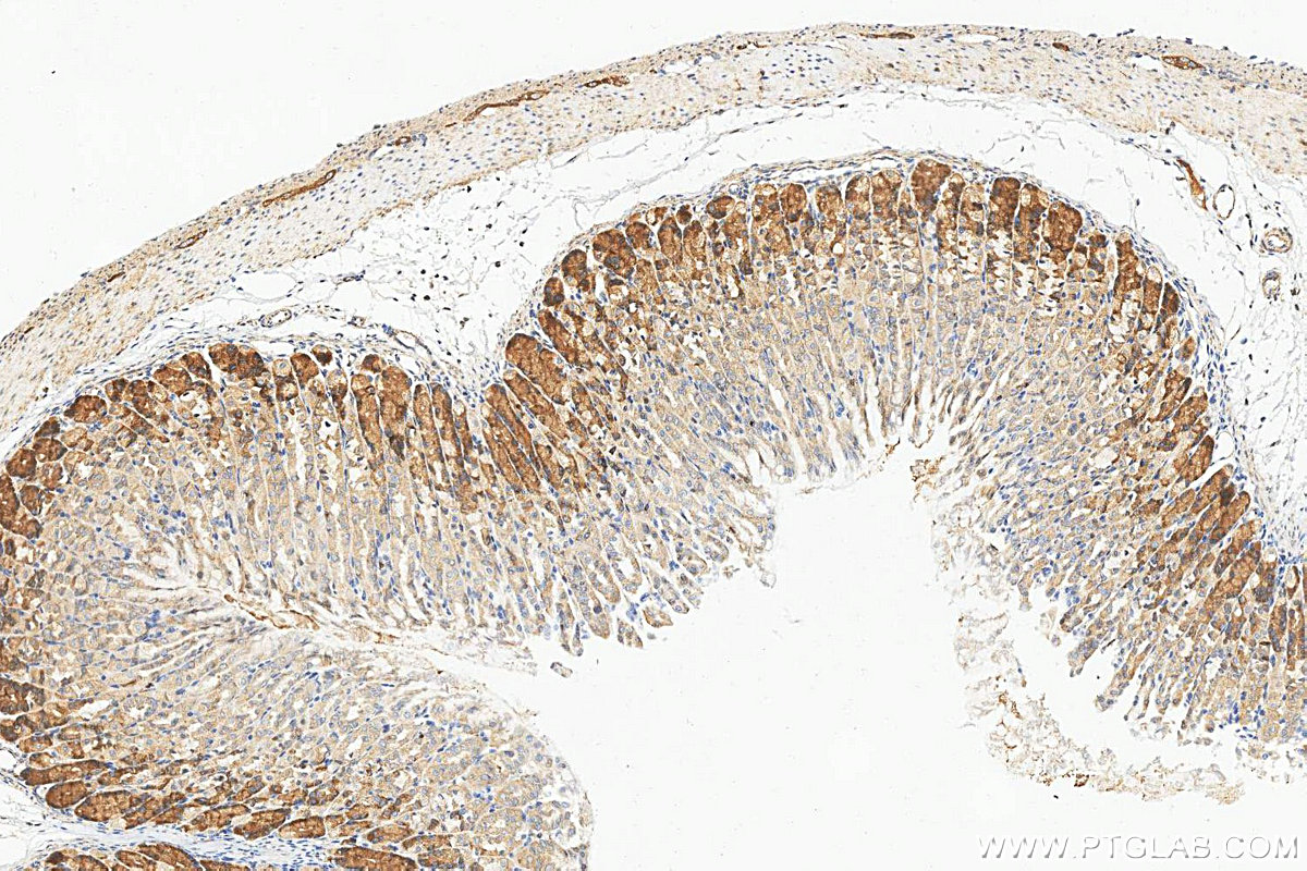
IHC staining of mouse stomach using 10582-1-AP (same clone as 10582-1-PBS)
Immunohistochemical analysis of paraffin-embedded mouse stomach tissue slide using 10582-1-AP (PELO antibody) at dilution of 1:500 (under 10x lens). Heat mediated antigen retrieval with Tris-EDTA buffer (pH 9.0). This data was developed using the same antibody clone with 10582-1-PBS in a different storage buffer formulation.
IHC staining of mouse stomach using 10582-1-AP (same clone as 10582-1-PBS)
Immunohistochemical analysis of paraffin-embedded mouse stomach tissue slide using 10582-1-AP (PELO antibody) at dilution of 1:500 (under 10x lens). Heat mediated antigen retrieval with Tris-EDTA buffer (pH 9.0). This data was developed using the same antibody clone with 10582-1-PBS in a different storage buffer formulation.
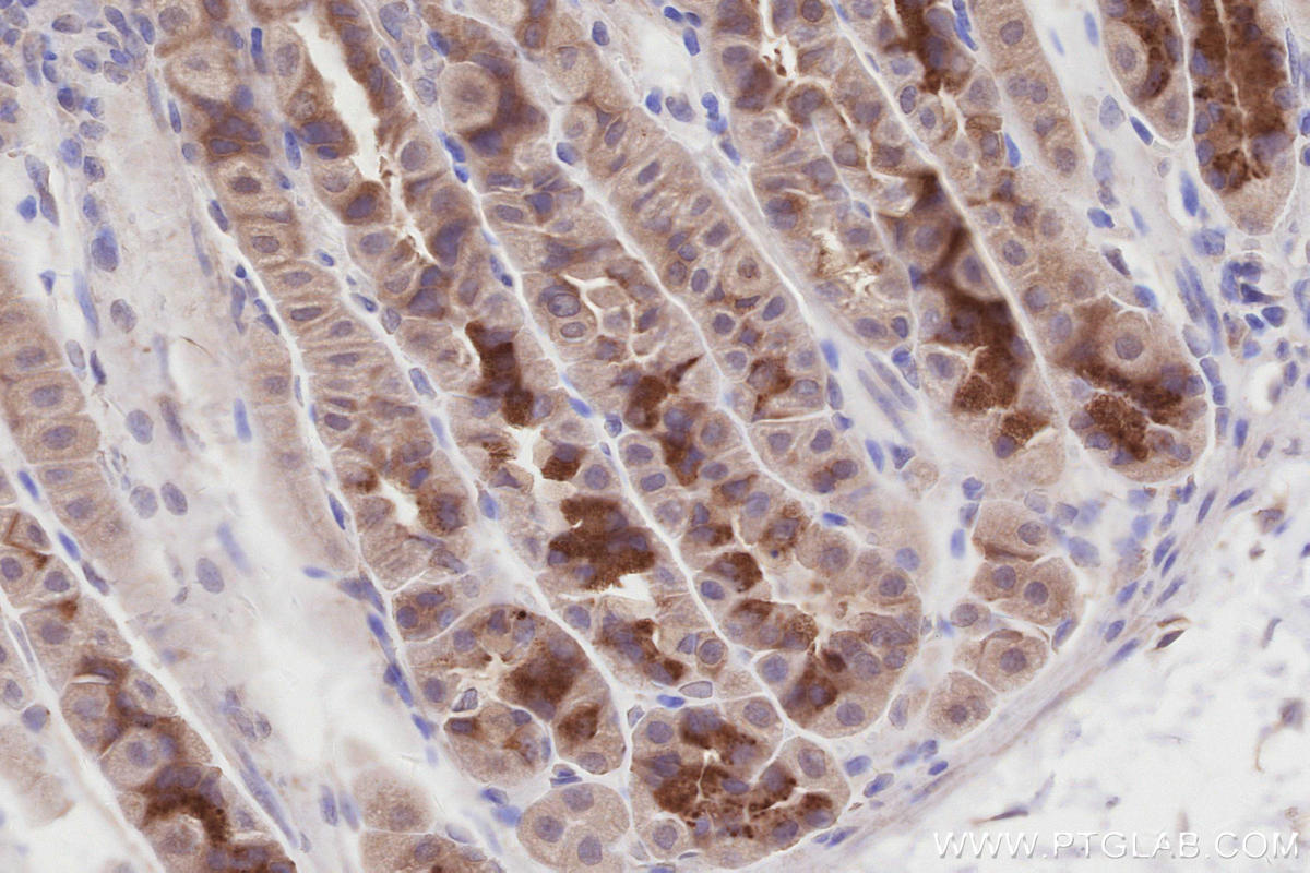
IHC staining of mouse stomach using 10582-1-AP (same clone as 10582-1-PBS)
Immunohistochemical analysis of paraffin-embedded mouse stomach tissue slide using 10582-1-AP (PELO antibody) at dilution of 1:1000 (under 40x lens). Heat mediated antigen retrieval with Tris-EDTA buffer (pH 9.0). This data was developed using the same antibody clone with 10582-1-PBS in a different storage buffer formulation.
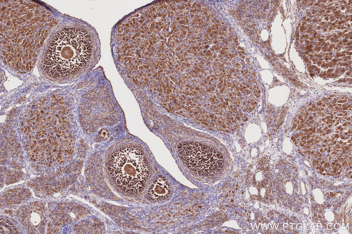
IHC staining of rat ovary using 10582-1-AP (same clone as 10582-1-PBS)
Immunohistochemical analysis of paraffin-embedded rat ovary tissue slide using 10582-1-AP (PELO antibody) at dilution of 1:500 (under 10x lens). Heat mediated antigen retrieval with Tris-EDTA buffer (pH 9.0). This data was developed using the same antibody clone with 10582-1-PBS in a different storage buffer formulation.
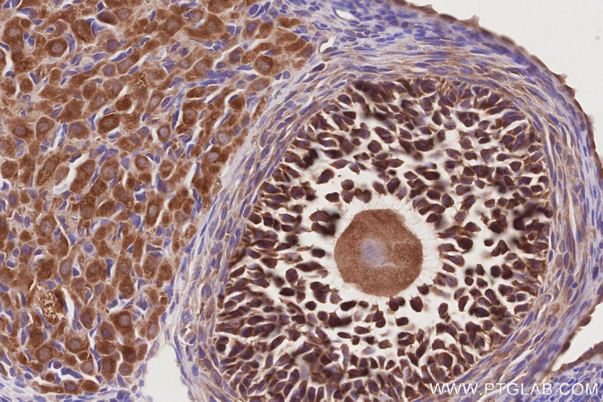
IHC staining of rat ovary using 10582-1-AP (same clone as 10582-1-PBS)
Immunohistochemical analysis of paraffin-embedded rat ovary tissue slide using 10582-1-AP (PELO antibody) at dilution of 1:500 (under 40x lens). Heat mediated antigen retrieval with Tris-EDTA buffer (pH 9.0). This data was developed using the same antibody clone with 10582-1-PBS in a different storage buffer formulation.
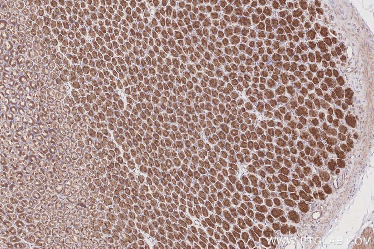
IHC staining of rat stomach using 10582-1-AP (same clone as 10582-1-PBS)
Immunohistochemical analysis of paraffin-embedded rat stomach tissue slide using 10582-1-AP (PELO antibody) at dilution of 1:500 (under 10x lens). Heat mediated antigen retrieval with Tris-EDTA buffer (pH 9.0). This data was developed using the same antibody clone with 10582-1-PBS in a different storage buffer formulation.