Use Able AI chat for product recommendations

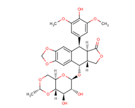
Etoposide

Etoposide (VP-16-213) 是一种拓扑异构酶 II 的抑制剂,通过与拓扑异构酶 II 和 DNA 形成复合物来抑制 DNA 合成 (IC50=60.3 μM)。Etoposide 具有抗肿瘤活性,可以诱导细胞凋亡、自噬。

CAS号

33419-42-0

分子式

C29H32O13

主要靶点

Autophagy|Apoptosis|Antibacterial|Antibiotic|Topoisomerase|Mitophagy

仅限科研使用

Cat No : CM00991

Print datasheet

Synonyms

依托泊甙|依托泊苷|prodrug|VP 16|VP16|VP-16|VP-16-213|P388|Topo II|Topoisomerase|leukemia|inhibit|Inhibitor|Mitochondrial Autophagy|p53|Mitophagy|HCT116|FBXW|Etoposide|chemotherapy|Bacterial|anti-cancer|Antibiotic|Autophagy|Apoptosis


产品信息

Etoposide (VP-16-213) is a topoisomerase II inhibitor that inhibits DNA synthesis by forming a complex with topoisomerase II and DNA (IC50=60.3 μM). Etoposide has antitumor activity and induces apoptosis and autophagy.

CAS号 33419-42-0
分子式 C29H32O13
主要靶点 Autophagy|Apoptosis|Antibacterial|Antibiotic|Topoisomerase|Mitophagy
主要通路 微生物学|DNA 损伤和修复|微生物学|凋亡|自噬|自噬
分子量 588.56
纯度 99.19%, 此纯度可做参考,具体纯度与批次有关系,可咨询客服
储存条件 Keep away from direct sunlight Powder: -20°C for 3 years | In solvent: -80°C for 1 year Shipping with blue ice/Shipping at ambient temperature.
别名 依托泊甙|依托泊苷|prodrug|VP 16|VP16|VP-16|VP-16-213|P388|Topo II|Topoisomerase|leukemia|inhibit|Inhibitor|Mitochondrial Autophagy|p53|Mitophagy|HCT116|FBXW|Etoposide|chemotherapy|Bacterial|anti-cancer|Antibiotic|Autophagy|Apoptosis

靶点活性

3LL CRL1642 cells viability:4 μM (48h,MTT assay)|A2058 cells viability:8.9 μM (24h)|A549 cells proliferation:0.32 μM (48h,SRB method)|A549 cells proliferation:0.4 μg/mL (72h,SRB assay)|COLO 205 cells proliferation:0.43 μM (48h,MTT assay)|CCRF-SB cells proliferation:0.1 μM (96h,MTT assay)|A2780 cells growth:0.07 μM (72h)|A2780 cells growth:1.3 μM (48h)|A375 cells proliferation:1.39 μM (48h,MTT assay)|A375 cells proliferation:0.5 μM (72h, MTT assay)|5637 cells proliferation:0.54 μM (96h,crystal violet assay)|CCRF-CEM cells viability:12.59 μM (72h,MTT assay)|Topo II:60.3 μM

体内活性

方法:为检测体内抗肿瘤活性,将 Etoposide (10 mg/kg) 和 Cisplatin (5-7.5 mg/kg) 腹腔注射给携带人子宫内膜腺癌肿瘤 Ishikawa 的 KSN nude 小鼠,每两天一次,持续两周。 结果:作为单一药物,Etoposide 对肿瘤生长几乎没有抑制作用。Etoposide 和 Cisplatin 联合治疗显著抑制肿瘤生长。[4] 方法:为检测体内抗肿瘤活性,将 Etoposide (80 mg/kg in 0.5% methylcellulose) 灌胃给药给携带人胶质母细胞肿瘤 U87 的免疫缺陷小鼠,每天一次,持续四十天。 结果:80 mg/kg Etoposide 抑制 U87 肿瘤生长,抑制率为95%。[5]

体外活性

方法:人宫颈癌细胞 HeLa 用 Etoposide (25-400 μM) 处理 24-48 h,使用 MTT 方法检测细胞活力。 结果:Etoposide 抑制 Hela 细胞增殖,处理 24 h 和 48 h 的 IC50 分别为 167.3 μM 和 52.7 μM。[1] 方法:人肺腺癌细胞 A549 用 Etoposide (0.75-3 μM) 处理 4 h,使用 Flow Cytometry 方法检测细胞周期情况。 结果:Etoposide 导致 G0/G1 和 S 期的 A549 细胞百分比显著降低。同时,G2/M 期的细胞显著增加。[2] 方法:小鼠胚胎成纤维细胞 MEFs 用 Etoposide (1.5-150 μM) 处理 3-18 h,使用 Western Blot 方法检测靶点蛋白表达水平。 结果:150 μM的 Etoposide 在 6 h 内诱导 Caspase-3 的强烈裂解,而 1.5 或 15 μM 仅在 18 h 后激活 Caspase-3。[3]

溶解度

DMSO:60.63 mg/mL (103.01 mM);10% DMSO+40% PEG300+5% Tween 80+45% Saline:5.89 mg/mL (10.01 mM)

细胞实验

After the Etoposide treatment, cells are removed from the dish with phosphate-buffered saline (PBS) containing 0.03% trypsin and 0.27 mM ethylenediaminetetraacetic acid (EDTA) and are diluted into culture dishes in appropriate numbers to yield between 20 and 200 colonies. After 12 days, cultures are fixed with methanol-acetic acid, stained with crystal violet, and scored for colonies containing more than 50 cells [5].

动物实验

The in vivo model for nude mice HB (NMHB) has been established. Only HB cells with embryonal components are grafted and reproduced successfully in this model. Each NMHB subsequently is transplanted into 50 mice for treatment groups. Treatment is initiated when the majority of the tumors reach a volume of 50-100 mm3. The mice are stratified according to their tumor volume and randomly assigned to groups of ten animals each. The animals injected with tumor are given ifosfamide, cisplatin, doxorubicin, etoposide (10 mg/kg/day, i.v.), and carboplatin as single agents in two blocks. One group of ten animals for each original xenograft served as a control group. After initiation of treatment, the tumor growth is recorded at 5-day intervals for 25-30 days and the relative tumor volumes are calculated. Twenty-four hours before the animals are sacrificed, bromodeoxyuridine (BrdU) is injected intraperitoneally for the semiquantitative determination of proliferation activity of the tumor cells (50 μg of BrdU/g body weight) [4].

参考文献

1.HGN ÇELEBİOĞLU, et al. Effects of thymoquinone and etoposide combination on cell viability and genotoxicity in human cervical cancer hela cells. Istanbul J Pharm. 2022; 52(3):258-264.
2.Weizhe Li, Hong-Yan Wang, Xiaolu Zhao, Hongguo Duan, Binghua Cheng, Yafei Liu, Mengjie Zhao et al. A methylation-phosphorylation switch determines Plk1 kinase activity and function in DNA damage repair [J]. Science Advances. 2019 Mar 6;5(3):eaau7566.
3.Litwiniec A, et al. Low-dose etoposide-treatment induces endoreplication and cell death accompanied by cytoskeletal alterations in A549 cells: Does the response involve senescence? The possible role of vimentin. Cancer Cell Int. 2013 Feb 5;13(1):9.
4.Jamil S, et al. Etoposide induces cell death via mitochondrial-dependent actions of p5Cancer Cell Int. 2015 Aug 7;15:79.
5.Suzuki M, et al. Anticancer activity of the combination of cisplatin and etoposide in endometrial cancer-bearing nude mice. Gynecol Oncol. 1991 Apr;41(1):41-5.
6.Panigrahy D, et al. Inhibition of tumor angiogenesis by oral etoposide. Exp Ther Med. 2010 Sep;1(5):739-746.
7.Lee KI, et al. Etoposide induces pancreatic β-cells cytotoxicity via the JNK/ERK/GSK-3 signaling-mediated mitochondria-dependent apoptosis pathway. Toxicol In Vitro. 2016 Jul 26. pii: S0887-2333(16)30147-3.
8.Calvani M, det al. Etoposide-Bevacizumab a new strategy against human melanoma cells expressing stem-like traits. Oncotarget. 2016 Jun 9. doi: 10.18632/oncotarget.9939.
9.Zhang J, Hirst A J, Duan F, et al. Darren Robinson, 3 Mark Jones, 2 Le Li, 4 Peizhe Wang, Peng Jiang, 4 Peter W. Andrews, 2 Ivana Barbaric, 2,* and Jie Na[J]. Anti-apoptotic Mutations Desensitize Human Pluripotent Stem Cells to Mitotic Stress and Enable Aneuploid Cell Survival. Stem Cell Reports.
10.Ruan C, Wang C, Gong X, et al. An integrative multi-omics approach uncovers the regulatory role of CDK7 and CDK4 in autophagy activation induced by silica nanoparticles[J]. Autophagy. 2020.

The molarity calculator equation

Mass (g) = Concentration (mol/L) × Volume (L) × Molecular Weight (g/mol)

质量   浓度   体积   分子量 *
=
×
×

The dilution calculator equation

Concentration (start) × Volume (start) = Concentration (final) × Volume (final)
This equation is commonly abbreviated as: C1V1 = C2V2

浓度 (start) × 体积 (start) = 浓度 (final) × 体积 (final)
×
=
×
C1   V1   C2   V2
||
New chat

Able

正在加载,请稍候...