验证数据展示


经过测试的应用
| Positive WB detected in | Raji cells, rat lung tissue, THP-1 cells |
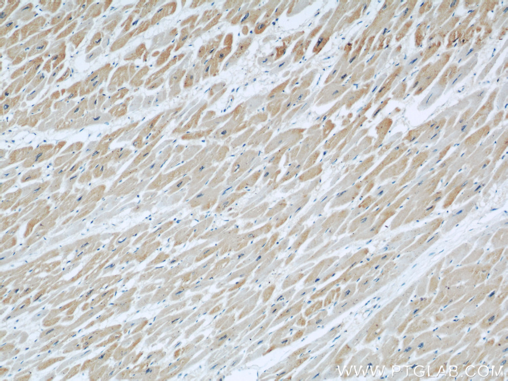
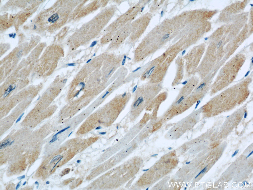
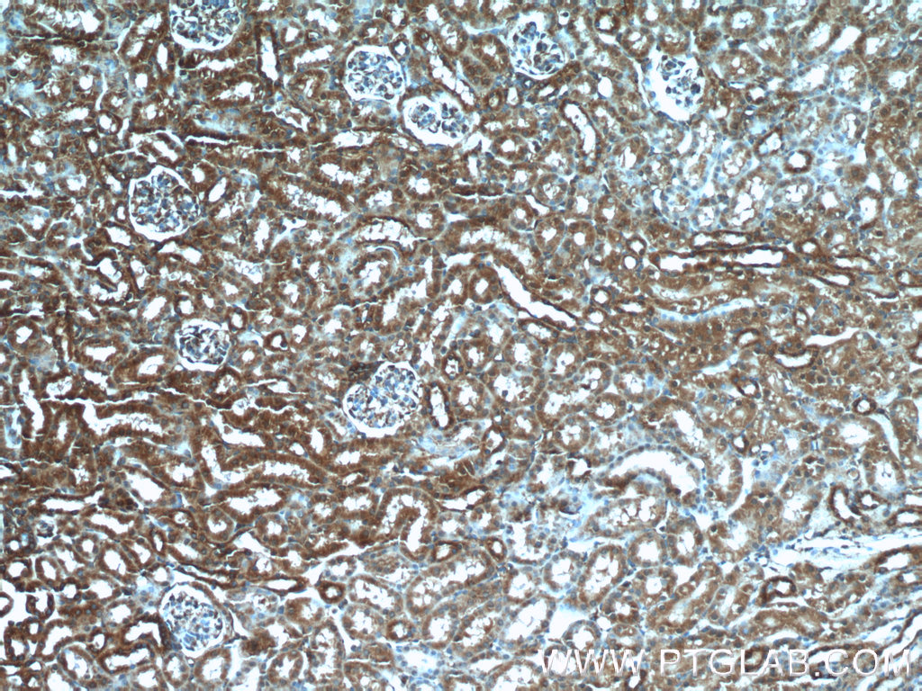
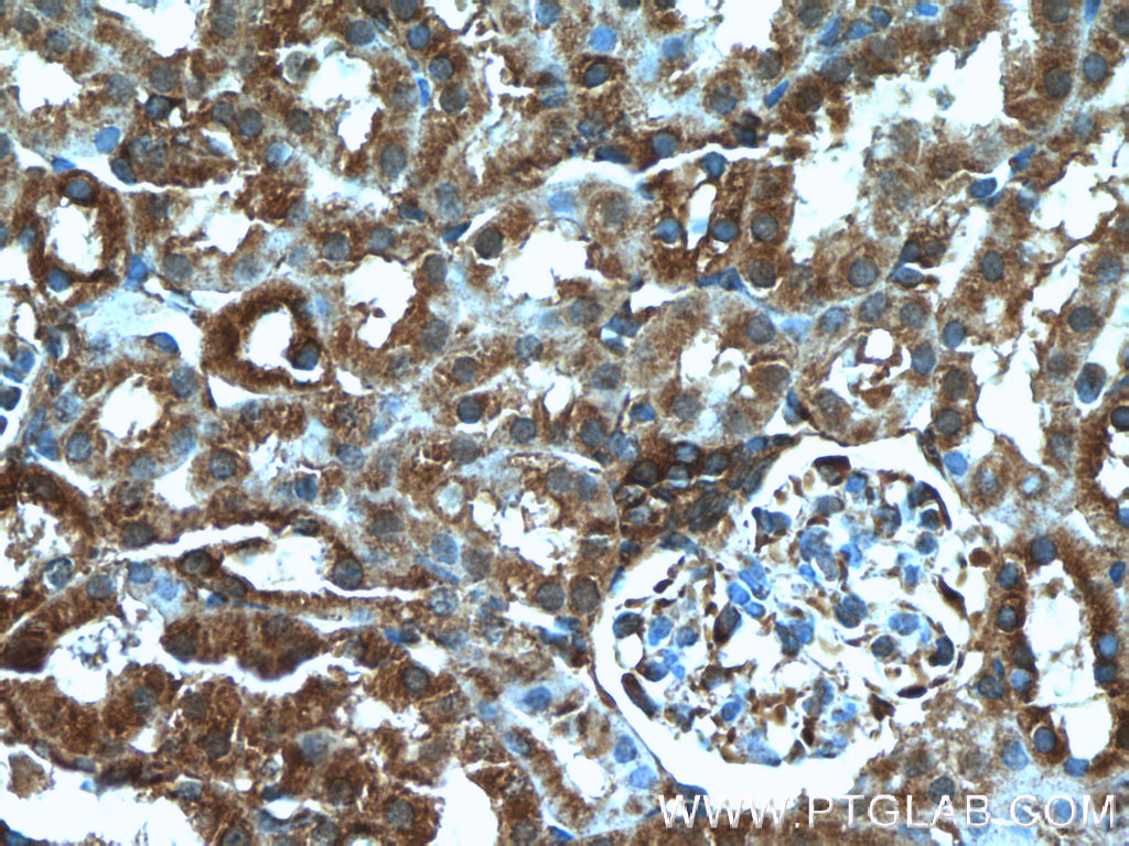
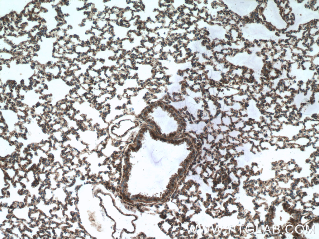
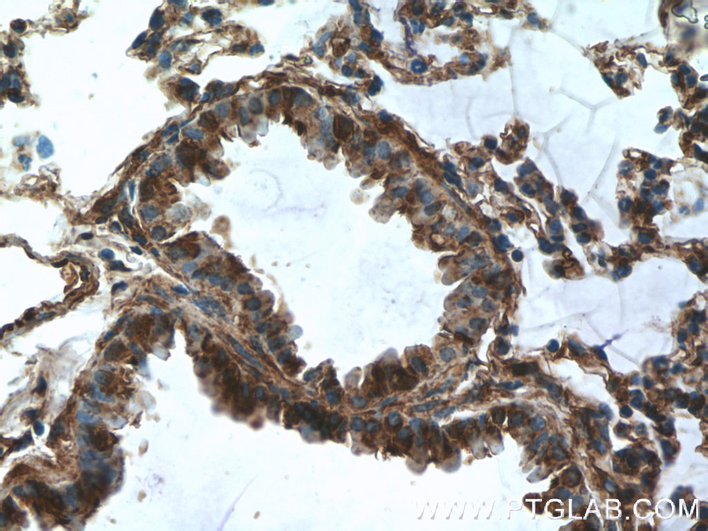
| Positive IHC detected in | rat lung tissue, human heart tissue, mouse kidney tissue, mouse lung tissue Note: suggested antigen retrieval with TE buffer pH 9.0; (*) Alternatively, antigen retrieval may be performed with citrate buffer pH 6.0 |
推荐稀释比
| 应用 | 推荐稀释比 |
|---|---|
| Western Blot (WB) | WB : 1:500-1:1000 |
| Immunohistochemistry (IHC) | IHC : 1:50-1:500 |
| It is recommended that this reagent should be titrated in each testing system to obtain optimal results. | |
| Sample-dependent, Check data in validation data gallery. | |
产品信息
11622-1-AP targets DOCK8 in WB, IHC, IF, IP, ELISA applications and shows reactivity with human, mouse, rat samples.
| 经测试应用 | WB, IHC, ELISA Application Description |
| 文献引用应用 | WB, IF, IP |
| 经测试反应性 | human, mouse, rat |
| 文献引用反应性 | human, mouse |
| 免疫原 |
CatNo: Ag2040 Product name: Recombinant human DOCK8 protein Source: e coli.-derived, PET28a Tag: 6*His Domain: 257-606 aa of BC019102 Sequence: EVYKLVIPILEAHREFRKLTLTHSKLQRAFDSIVNKDHKRMFGTYFRVGFFGSKFGDLDEQEFVYKEPAITKLPEISHRLEAFYGQCFGAEFVEVIKDSTPVDKTKLDPNKAYIQITFVEPYFDEYEMKDRVTYFEKNFNLRRFMYTTPFTLEGRPRGELHEQYRRNTVLTTMHAFPYIKTRISVIQKEEFVLTPIEVAIEDMKKKTLQLAVAINQEPPDAKMLQMVLQGSVGATVNQGPLEVAQVFLAEIPADPKLYRHHNKLRLCFKEFIMRCGEAVEKNKRLITADQREYQQELKKNYNKLKENLRPMIERKIPELYKPIFRVESQKRDSFHRSSFRKCETQLSQGS 种属同源性预测 |
| 宿主/亚型 | Rabbit / IgG |
| 抗体类别 | Polyclonal |
| 产品类型 | Antibody |
| 全称 | dedicator of cytokinesis 8 |
| 别名 | dedicator of cytokinesis 8, MRD2 |
| 计算分子量 | 2031 aa, 231 kDa |
| 观测分子量 | 230-239 kDa |
| GenBank蛋白编号 | BC019102 |
| 基因名称 | DOCK8 |
| Gene ID (NCBI) | 81704 |
| RRID | AB_10216360 |
| 偶联类型 | Unconjugated |
| 形式 | Liquid |
| 纯化方式 | Antigen affinity purification |
| UNIPROT ID | Q8NF50 |
| 储存缓冲液 | PBS with 0.02% sodium azide and 50% glycerol, pH 7.3. |
| 储存条件 | Store at -20°C. Stable for one year after shipment. Aliquoting is unnecessary for -20oC storage. |
背景介绍
Background
Dedicator of Cytokinesis 8 (DOCK8) is a protein that regulates the actin cytoskeleton, with particular importance in immune cells and a key role in innate and adaptive immune responses.
What is the molecular weight of DOCK8?
231 kDa. DOCK8 is a protein composed of 2031 amino acids and is a guanine nucleotide exchange factor (GEF).
What is the function of DOCK8?
DOCK8 is a member of the DOCK family of proteins, which have a unique DRH2 domain enabling them to act as GEFs and so controlling a range of cellular processes in various signaling pathways (PMID: 12432077). The specific target of DOCK8 is Cell division control protein 42 homolog (Cdc42), a small GTPase that is involved in regulation of the cell cycle and forms a complex. DOCK8 also acts as a scaffold molecule in this complex that initiates actin polymerization via the Wiskott-Aldrich Syndrome protein (WASp) (PubMed: 28028151, PubMed: 22461490).
What diseases are associated with DOCK8?
The role of DOCK8 in immunity was first identified with the study of DOCK8-deficient patients who presented with combined immunodeficiency (PMID: 19776401; PMID: 20004785). The subsequent study of DOCK8 in immune cells such as T cells, natural killer (NK) cells, and B cells has revealed how it regulates their normal function. This includes the regulation of immune synapse formation, immune cell trafficking, regulation of dendritic cell polarization, and cytokine production (PMID: 28366940).
DOCK8 deficiency is caused by a number of different mutations in the gene. It leads to the autosomal recessive form of the immunodeficiency disease Hyper-IgE syndrome, or Job's syndrome. The symptoms of DOCK8 deficiency include eczema, high levels of serum IgE, hypereosinophilia, and recurrent respiratory and skin infections as a result of impaired immune cell function.
实验方案
| Product Specific Protocols | |
|---|---|
| IHC protocol for DOCK8 antibody 11622-1-AP | Download protocol |
| WB protocol for DOCK8 antibody 11622-1-AP | Download protocol |
| Standard Protocols | |
|---|---|
| Click here to view our Standard Protocols |
发表文章
| Species | Application | Title |
|---|---|---|
Nat Immunol Heme drives hemolysis-induced susceptibility to infection via disruption of phagocyte functions.
| ||
Cell Death Differ CCL2 regulation of MST1-mTOR-STAT1 signaling axis controls BCR signaling and B-cell differentiation. | ||
Diabetes Dedicator of Cytokinesis 5 Regulates Keratinocyte Function and Promotes Diabetic Wound Healing. | ||
Oncotarget CD147 promotes Src-dependent activation of Rac1 signaling through STAT3/DOCK8 during the motility of hepatocellular carcinoma cells.
| ||
iScience DOCK8-expressing T follicular helper cells newly generated beyond self-organized criticality cause systemic lupus erythematosus.
| ||
J Alzheimers Dis A novel finding relates to the involvement of ATF3/DOCK8 in Alzheimer's disease pathogenesis |