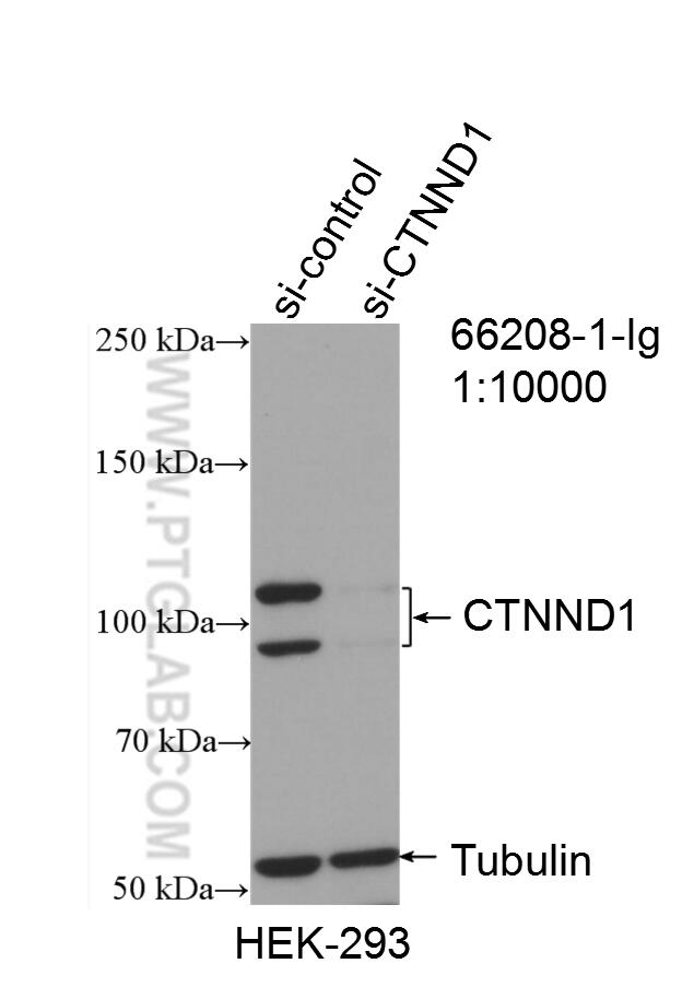
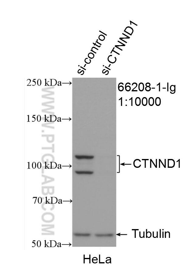
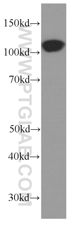
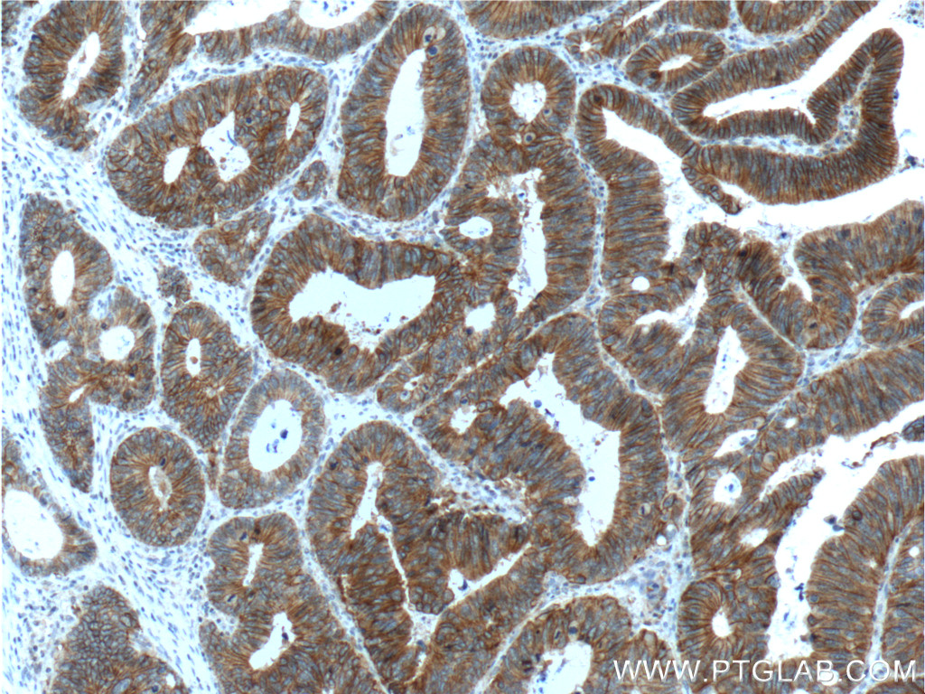
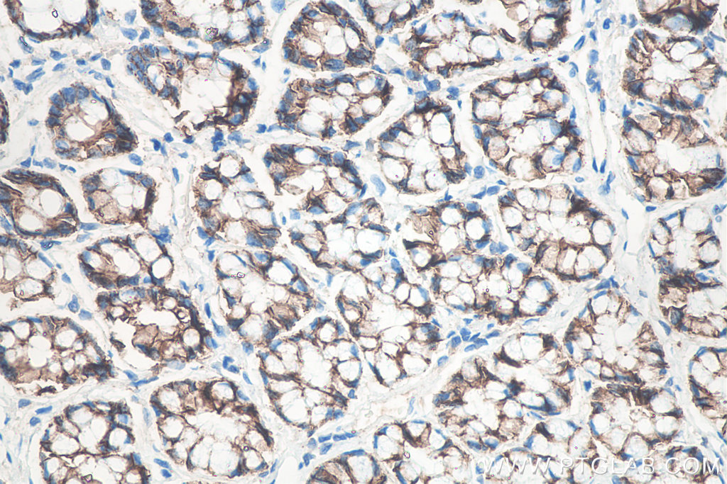
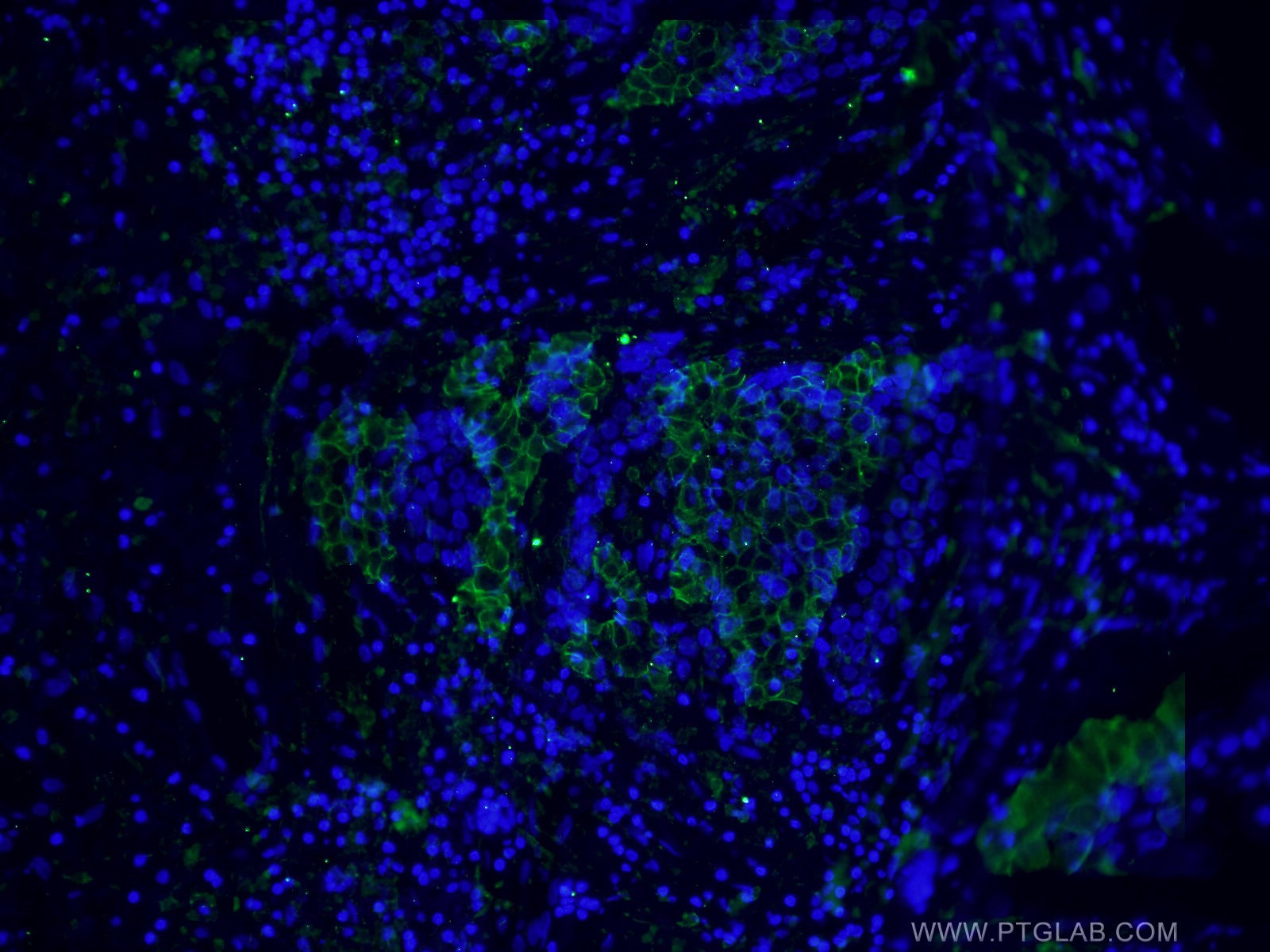
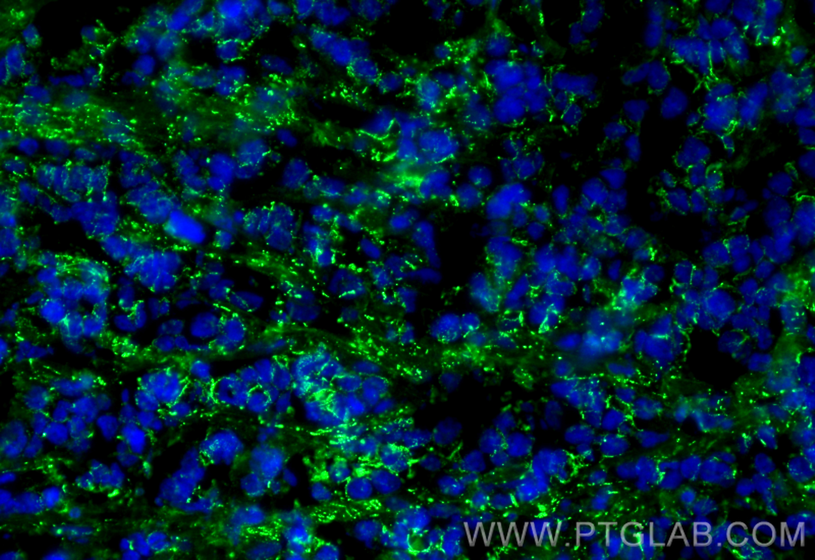
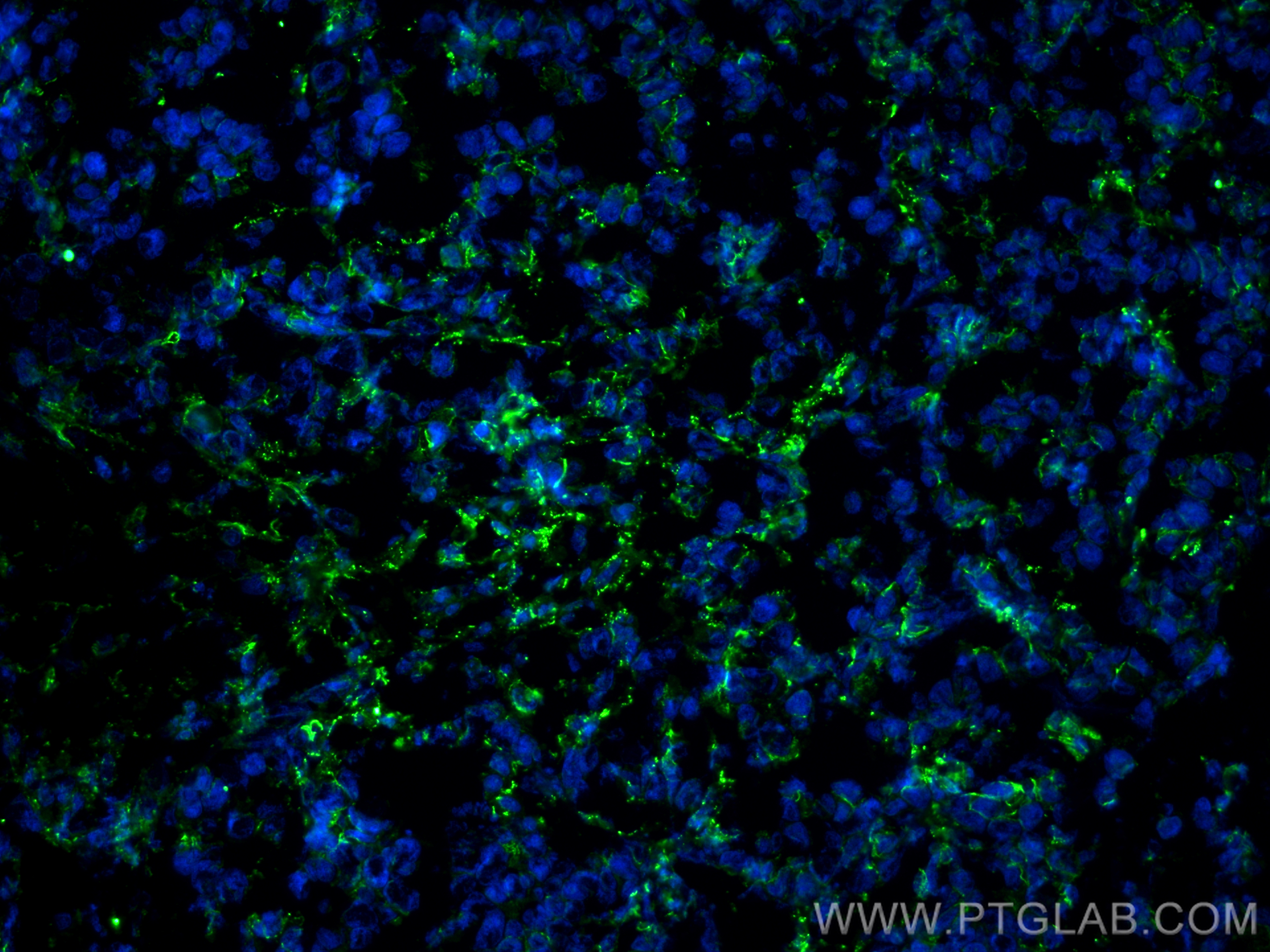
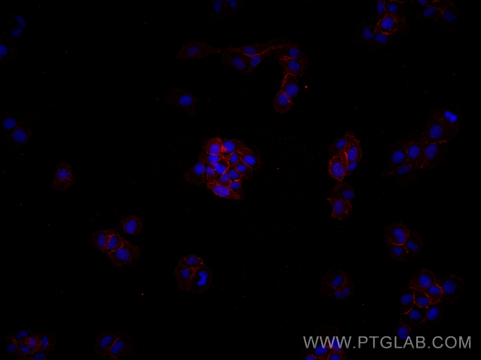
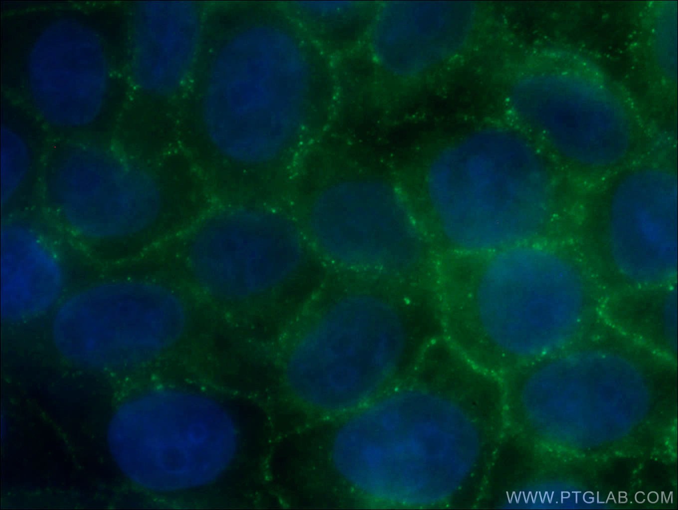
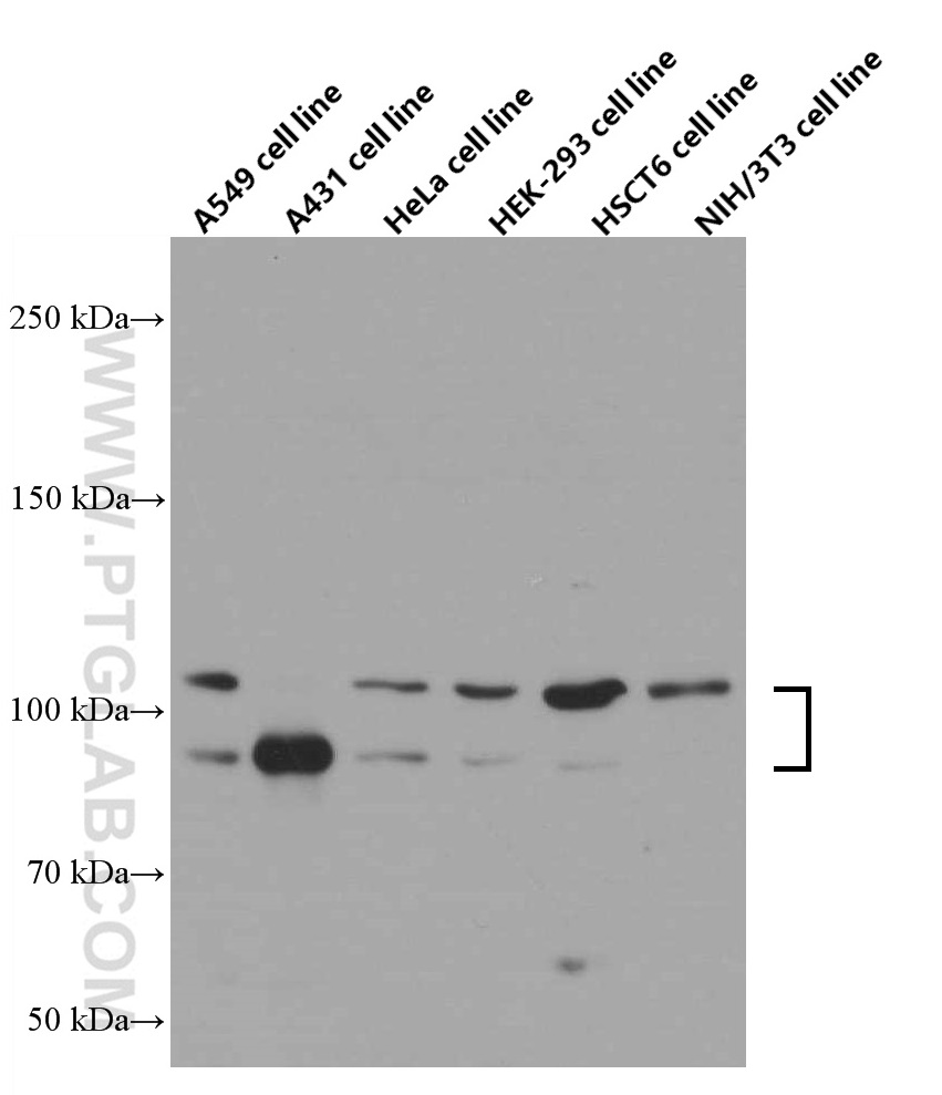
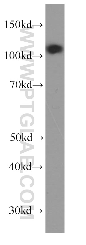
验证数据展示
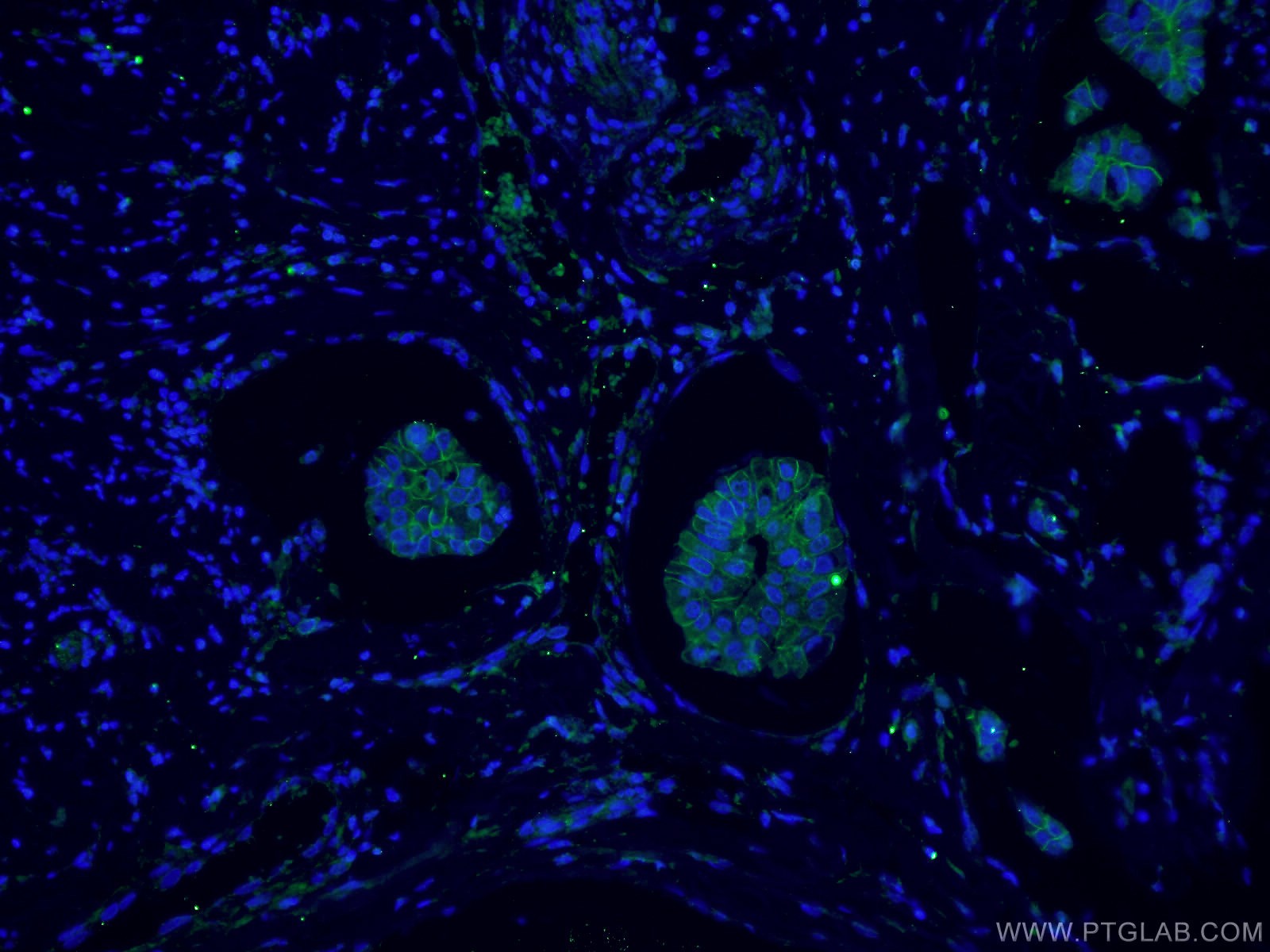

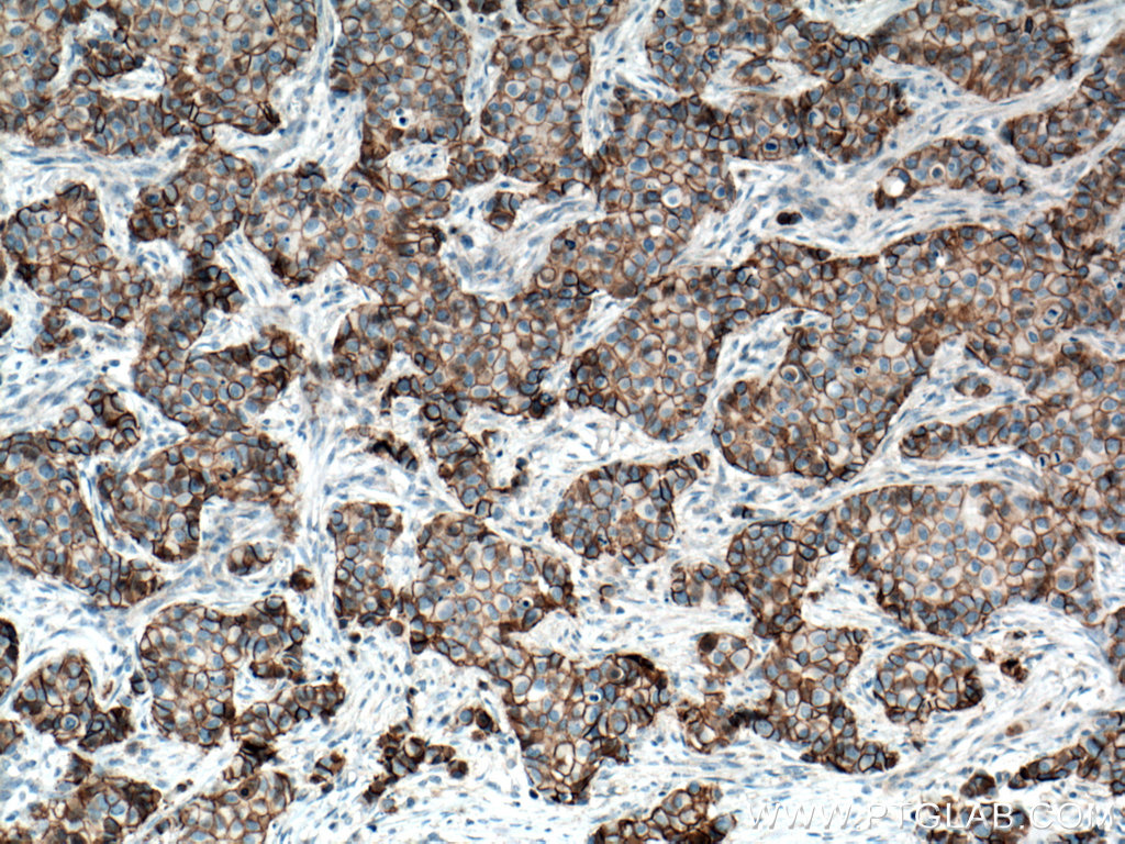
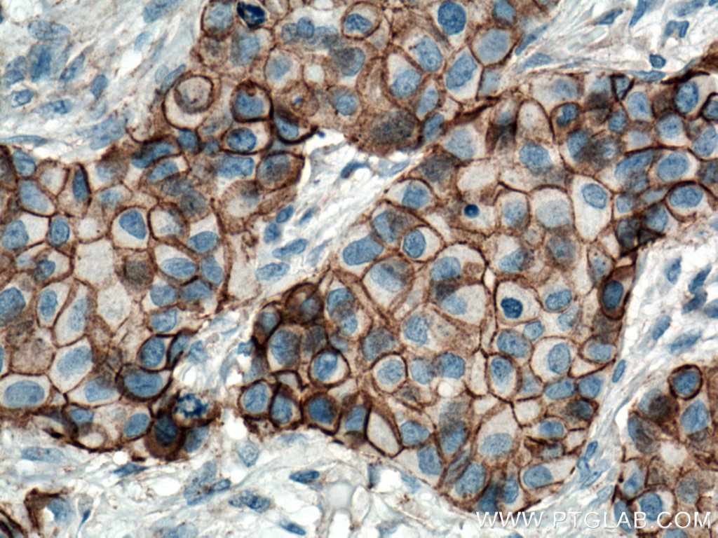

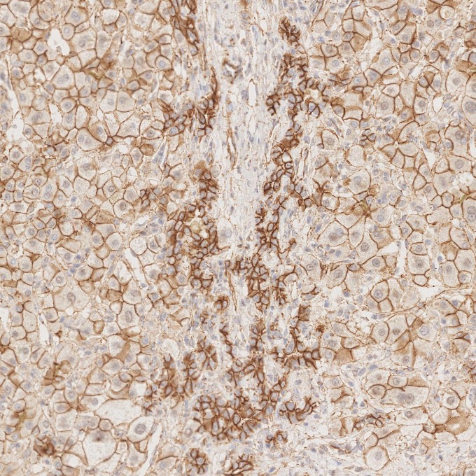
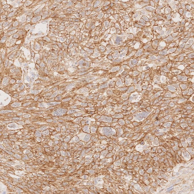
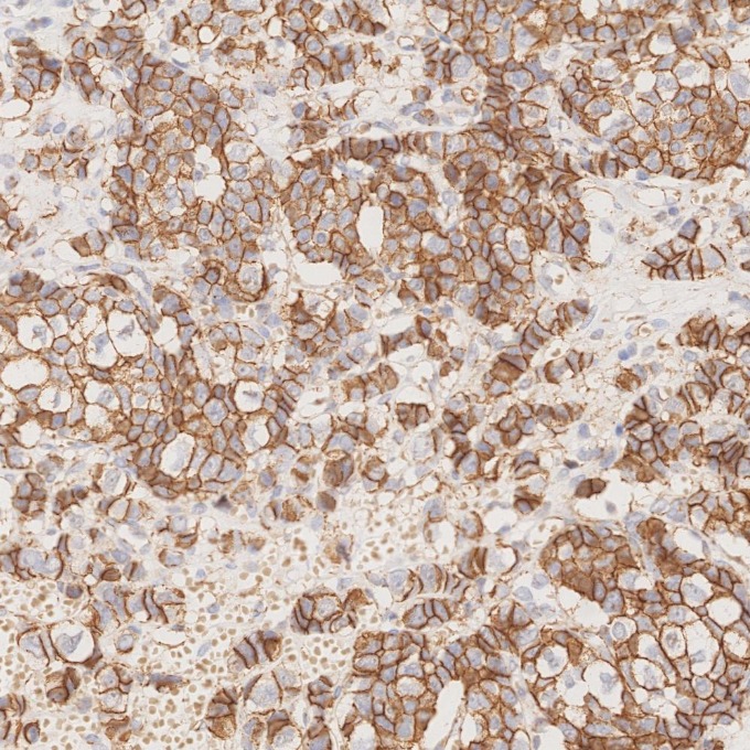
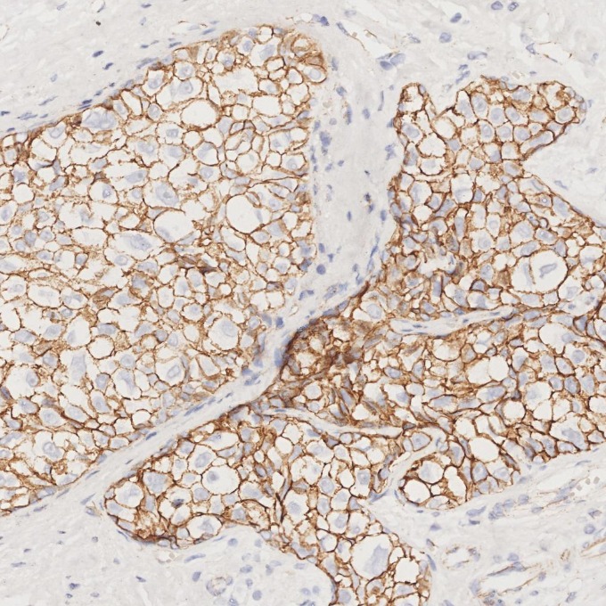
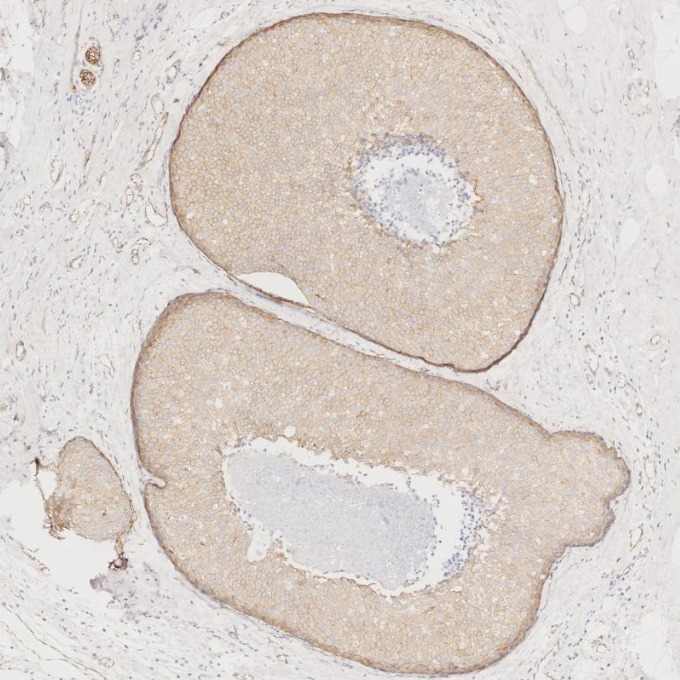
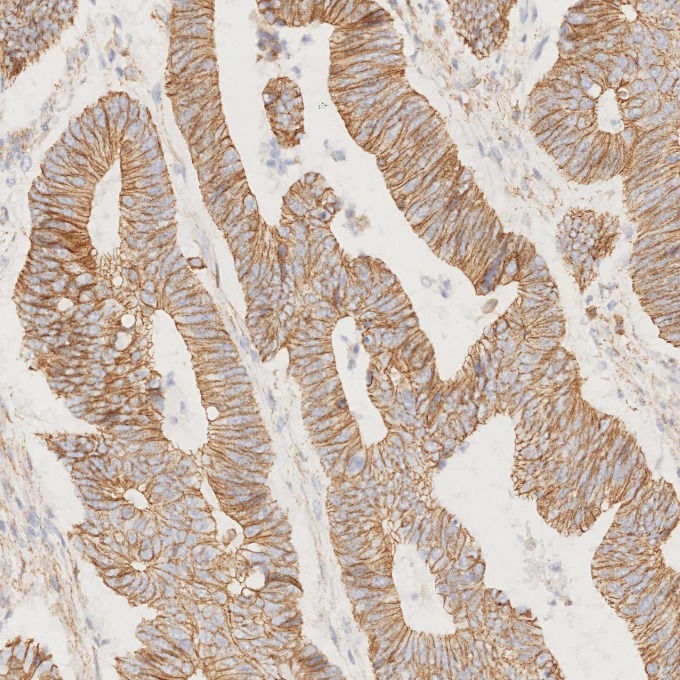
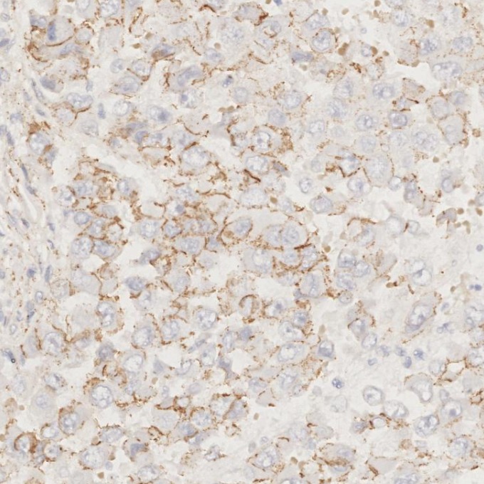
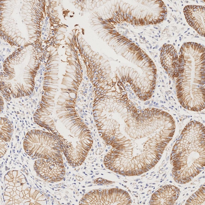
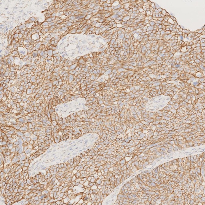
经过测试的应用
| Positive WB detected in | A549 cells, fetal human brain tissue, HEK-293 cells, HeLa cells, mouse brain tissue, A431 cells, HSC-T6 cells, NIH/3T3 cells |
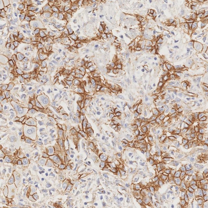
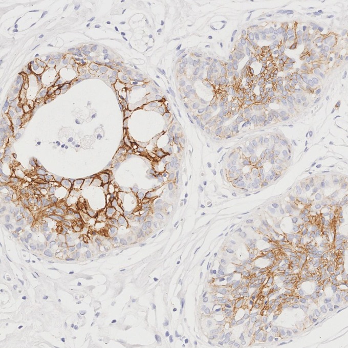
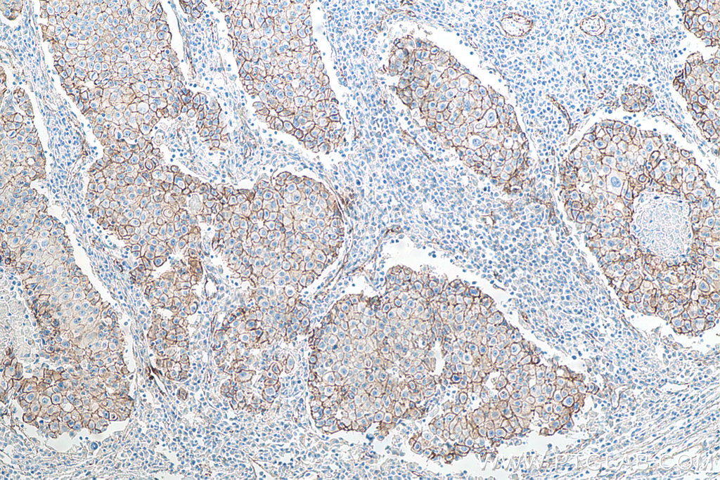
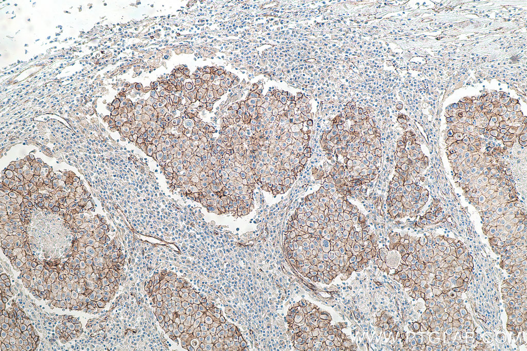
| Positive IHC-Autostainer detected in | human ovary cancer tissue, human stomach cancer tissue, human breast cancer tissue, human liver tissue |
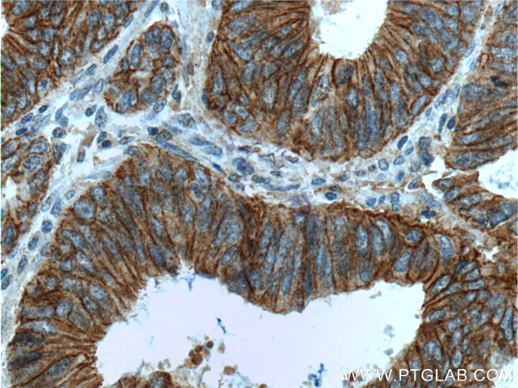
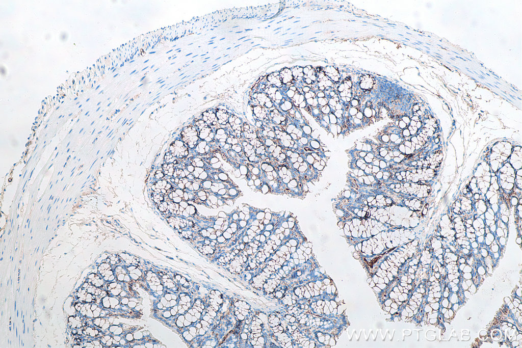
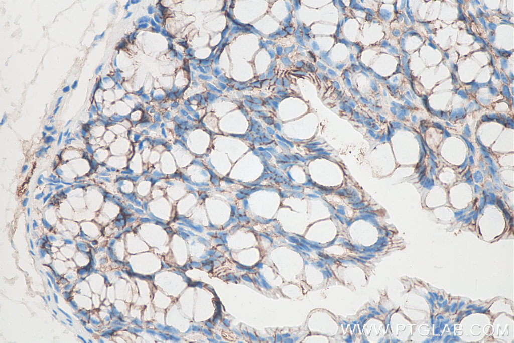
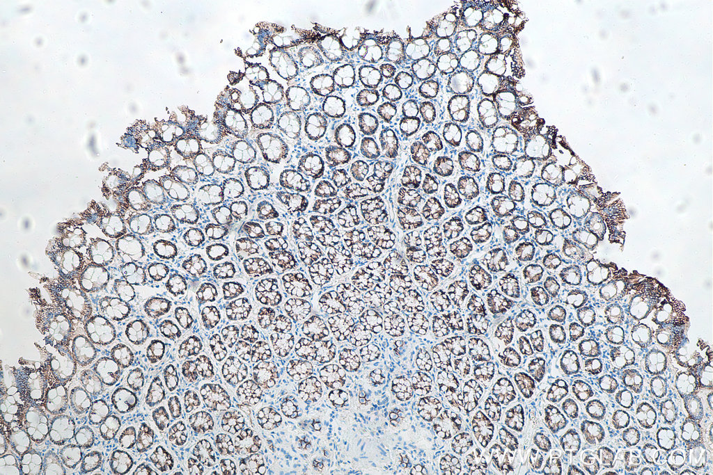
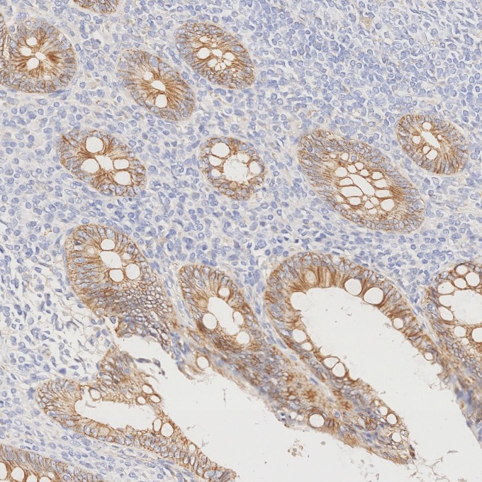
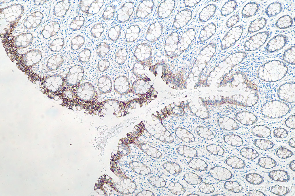
| Positive IHC detected in | human breast cancer tissue, human liver tissue, human breast tissue, human stomach cancer tissue, human colon tissue, human ovary cancer tissue, mouse colon tissue, rat colon tissue, human appendicitis tissue, human colon cancer tissue, human colorectal cancer tissue Note: suggested antigen retrieval with TE buffer pH 9.0; (*) Alternatively, antigen retrieval may be performed with citrate buffer pH 6.0 |
| Positive IF/ICC detected in | MCF-7 cells, HeLa cells, A431 cells |
推荐稀释比
| 应用 | 推荐稀释比 |
|---|---|
| Western Blot (WB) | WB : 1:2000-1:10000 |
| Immunohistochemistry (IHC)-AUTOSTAINER | IHC-AUTOSTAINER : 1:2500-1:10000 |
| Immunohistochemistry (IHC) | IHC : 1:250-1:1000 |
| Immunofluorescence (IF)/ICC | IF/ICC : 1:200-1:800 |
| It is recommended that this reagent should be titrated in each testing system to obtain optimal results. | |
| Sample-dependent, Check data in validation data gallery. | |
产品信息
66208-1-Ig targets p120 Catenin in WB, IHC, IHC-Autostainer, IF/ICC, IP, ELISA applications and shows reactivity with human, mouse, rat samples.
| 经测试应用 | WB, IHC, IHC-Autostainer, IF/ICC, ELISA Application Description |
| 文献引用应用 | WB, IF, IP |
| 经测试反应性 | human, mouse, rat |
| 文献引用反应性 | human, mouse, rat, canine |
| 免疫原 |
CatNo: Ag2824 Product name: Recombinant human CTNND1 protein Source: e coli.-derived, PGEX-4T Tag: GST Domain: 546-830 aa of BC010501 Sequence: GYELLFQPEVVRIYISLLKESKTPAILEASAGAIQNLCAGRWTYGRYIRSALRQEKALSAIADLLTNEHERVVKAASGALRNLAVDARNKELIGKHAIPNLVKNLPGGQQNSSWNFSEDTVISILNTINEVIAENLEAAKKLRETQGIEKLVLINKSGNRSEKEVRAAALVLQTIWGYKELRKPLEKEGWKKSDFQVNLNNASRSQSSHSYDDSTLPLIDRNQKSDKKPDREEIQMSNMGSNTKSLDNNYSTPNERGDHNKTLDRSGDLGDMEPLKGTTPLMQKI 种属同源性预测 |
| 宿主/亚型 | Mouse / IgG2b |
| 抗体类别 | Monoclonal |
| 产品类型 | Antibody |
| 全称 | catenin (cadherin-associated protein), delta 1 |
| 别名 | CTNND1, Cadherin-associated Src substrate, CAS, Catenin delta 1, Catenin delta-1 |
| 计算分子量 | 948 aa, 105 kDa |
| 观测分子量 | 90-120 kDa |
| GenBank蛋白编号 | BC010501 |
| 基因名称 | p120 Catenin |
| Gene ID (NCBI) | 1500 |
| RRID | AB_2881599 |
| 偶联类型 | Unconjugated |
| 形式 | Liquid |
| 纯化方式 | Protein A purification |
| UNIPROT ID | O60716 |
| 储存缓冲液 | PBS with 0.02% sodium azide and 50% glycerol, pH 7.3. |
| 储存条件 | Store at -20°C. Stable for one year after shipment. Aliquoting is unnecessary for -20oC storage. |
背景介绍
Catenins were discovered as proteins that are linked to the cytoplasmic domain of transmembrane cadherins (PMID: 9653641). p120 catenin, also called p120 ctn or catenin delta-1, regulates cell-cell adhesion through its interaction with the cytoplasmic tail of classical and type II cadherins. p120 catenin is a tyrosine kinase substrate implicated in cell transformation by SRC, as well as in ligand-induced receptor signaling through the EGF receptor, the PDGF receptor, and the CSF1 receptor. Different expression patterns of p120 catenin in lobular and ductal carcinomas of breast have been reported: membrane stain for ductal carcinoma and cytoplasmic stain for lobular carcinoma (PMID: 24966968). Different isoforms of p120 catenin are variably expressed in different tissues as a result of alternative splicing and the use of multiple translation initiation codons (PMID: 19150613).
实验方案
| Product Specific Protocols | |
|---|---|
| IF protocol for p120 Catenin antibody 66208-1-Ig | Download protocol |
| IHC protocol for p120 Catenin antibody 66208-1-Ig | Download protocol |
| WB protocol for p120 Catenin antibody 66208-1-Ig | Download protocol |
| Standard Protocols | |
|---|---|
| Click here to view our Standard Protocols |
发表文章
| Species | Application | Title |
|---|---|---|
Cancer Discov Spatial Transcriptomics of Intraductal Papillary Mucinous Neoplasms of the Pancreas Identifies NKX6-2 as a Driver of Gastric Differentiation and Indolent Biological Potential | ||
Proc Natl Acad Sci U S A SENCR stabilizes vascular endothelial cell adherens junctions through interaction with CKAP4. | ||
Biomed Pharmacother Inhibition of autophagy and RIP1/RIP3/MLKL-mediated necroptosis by edaravone attenuates blood spinal cord barrier disruption following spinal cord injury | ||
Cell Mol Life Sci Distinct VE-cadherin serine and tyrosine phosphorylation sites and their role for inflammation-induced vascular permeability in vivo | ||
Mol Oncol Kaiso phosphorylation at threonine 606 leads to its accumulation in the cytoplasm, reducing its transcriptional repression of the tumour suppressor CDH1. | ||
Development Ontogenesis of the tear drainage system requires Prickle1-driven polarized basement membrane deposition. |