WB Figures
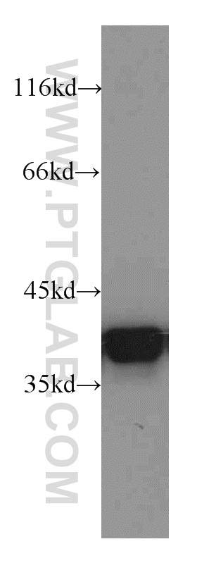
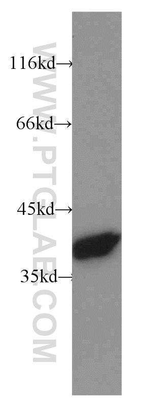
WB analysis of A431 using 21990-1-AP (same clone as 21990-1-PBS)
A431 cells were subjected to SDS PAGE followed by western blot with 21990-1-AP (Annexin A1 antibody) at dilution of 1:2000 incubated at room temperature for 1.5 hours. This data was developed using the same antibody clone with 21990-1-PBS in a different storage buffer formulation.
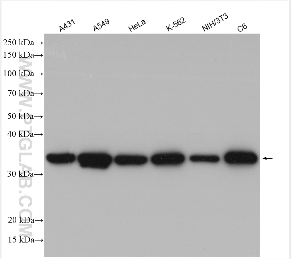
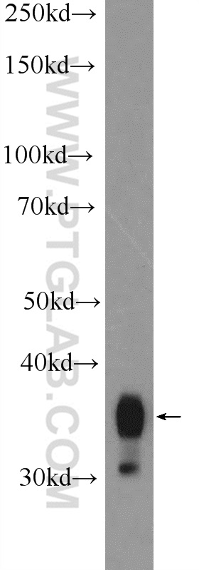
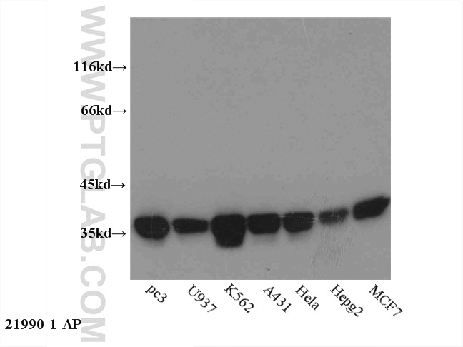
WB analysis using 21990-1-AP (same clone as 21990-1-PBS)
Various lysates were subjected to SDS PAGE followed by western blot with 21990-1-AP (Annexin A1 antibody) at dilution of 1:25000 incubated at room temperature for 1.5 hours. This data was developed using the same antibody clone with 21990-1-PBS in a different storage buffer formulation.
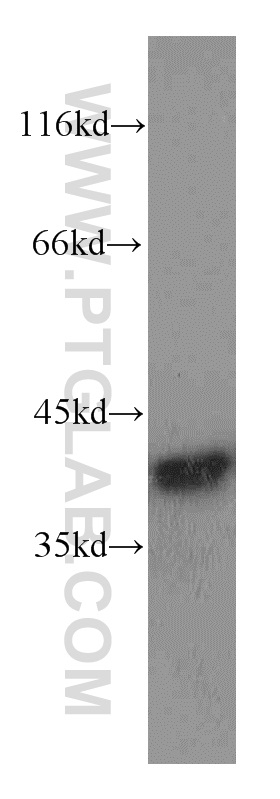
WB analysis using 21990-1-AP (same clone as 21990-1-PBS)
A549 cells were subjected to SDS PAGE followed by western blot with 21990-1-AP (Annexin A1 antibody) at dilution of 1:40000 incubated at room temperature for 1.5 hours. This data was developed using the same antibody clone with 21990-1-PBS in a different storage buffer formulation.
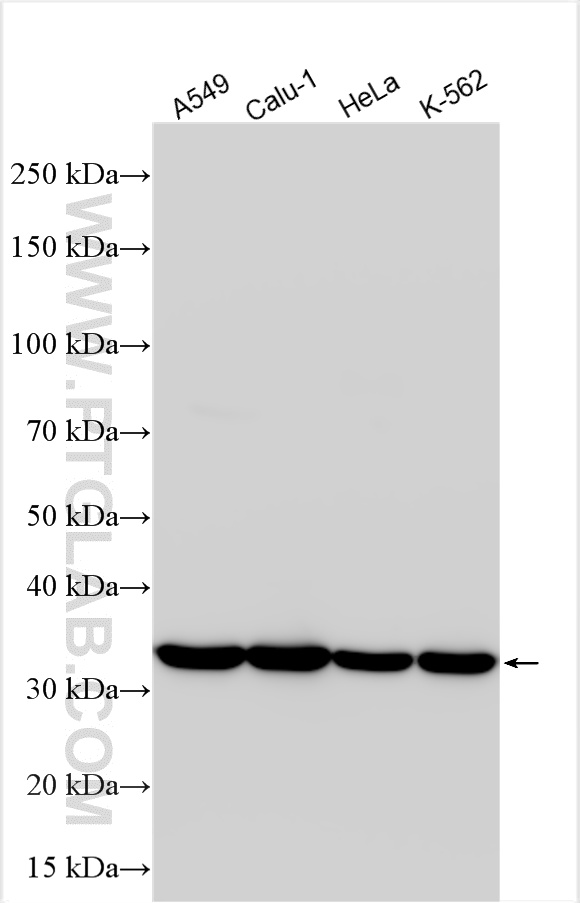
WB analysis using 21990-1-AP (same clone as 21990-1-PBS)
Various lysates were subjected to SDS PAGE followed by western blot with 21990-1-AP (Annexin A1 antibody) at dilution of 1:60000 incubated at room temperature for 1.5 hours. This data was developed using the same antibody clone with 21990-1-PBS in a different storage buffer formulation.
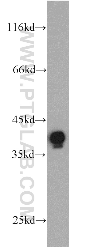
WB analysis of C6 using 21990-1-AP (same clone as 21990-1-PBS)
C6 cells were subjected to SDS PAGE followed by western blot with 21990-1-AP (Annexin A1 antibody at dilution of 1:2000 incubated at room temperature for 1.5 hours. This data was developed using the same antibody clone with 21990-1-PBS in a different storage buffer formulation.
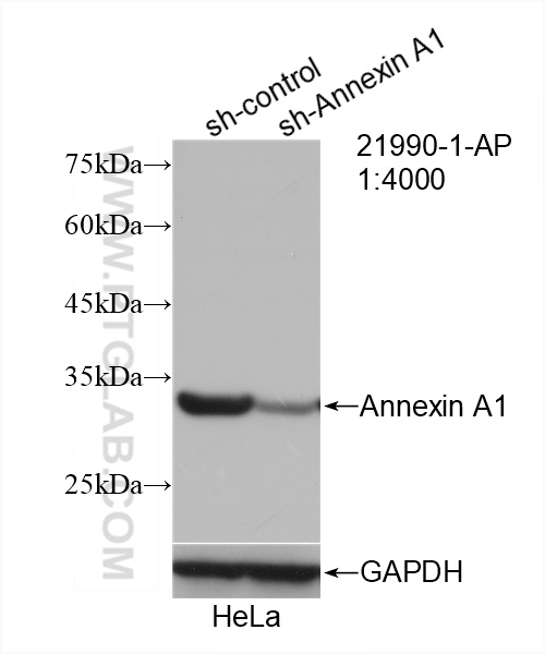
WB analysis of HeLa using 21990-1-AP (same clone as 21990-1-PBS)
WB result of Annexin A1 antibody (21990-1-AP; 1:4000; incubated at room temperature for 1.5 hours) with sh-Control and sh-Annexin A1 transfected HeLa cells. This data was developed using the same antibody clone with 21990-1-PBS in a different storage buffer formulation.
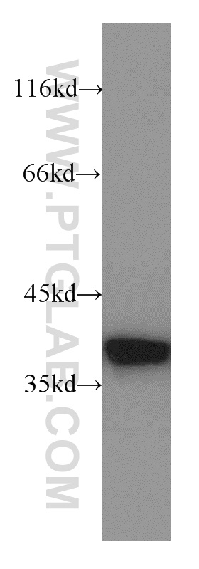
WB analysis of HepG2 using 21990-1-AP (same clone as 21990-1-PBS)
HepG2 cells were subjected to SDS PAGE followed by western blot with 21990-1-AP (Annexin A1 antibody) at dilution of 1:2000 incubated at room temperature for 1.5 hours. This data was developed using the same antibody clone with 21990-1-PBS in a different storage buffer formulation.
WB analysis of MCF-7 using 21990-1-AP (same clone as 21990-1-PBS)
MCF7 cells were subjected to SDS PAGE followed by western blot with 21990-1-AP (Annexin A1 antibody) at dilution of 1:2000 incubated at room temperature for 1.5 hours. This data was developed using the same antibody clone with 21990-1-PBS in a different storage buffer formulation.
WB analysis of MDA-MB-453s using 21990-1-AP (same clone as 21990-1-PBS)
MDA-MB-453s cells were subjected to SDS PAGE followed by western blot with 21990-1-AP (Annexin A1 antibody) at dilution of 1:1000 incubated at room temperature for 1.5 hours. This data was developed using the same antibody clone with 21990-1-PBS in a different storage buffer formulation.
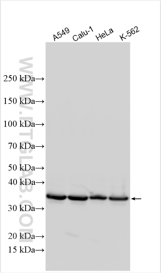
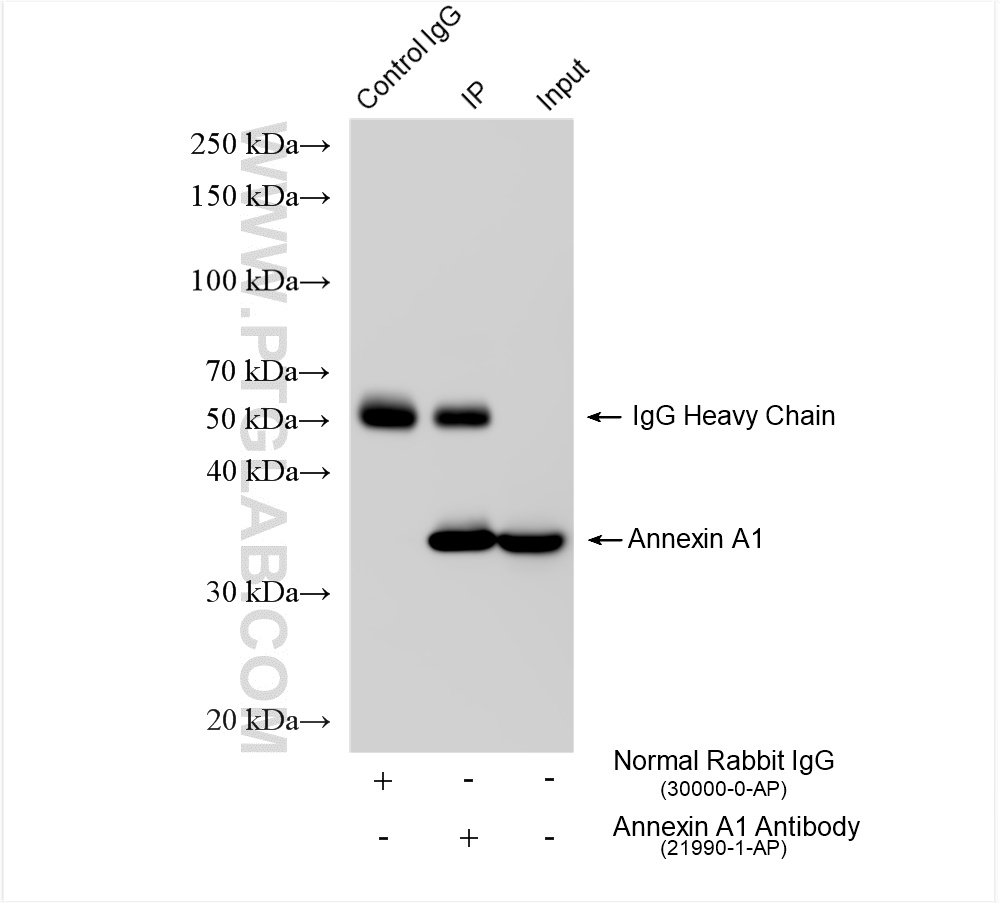
WB analysis of multi-cells using 21990-1-AP (same clone as 21990-1-PBS)
WB result of 21990-1-AP (Annexin A1 antibody). This data was developed using the same antibody clone with 21990-1-PBS in a different storage buffer formulation.
WB analysis of U-937 using 21990-1-AP (same clone as 21990-1-PBS)
U-937 cells were subjected to SDS PAGE followed by western blot with 21990-1-AP (Annexin A1 antibody) at dilution of 1:2000 incubated at room temperature for 1.5 hours. This data was developed using the same antibody clone with 21990-1-PBS in a different storage buffer formulation.
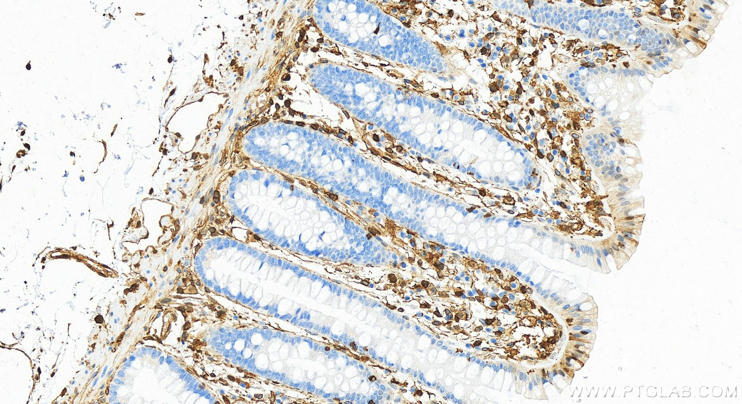
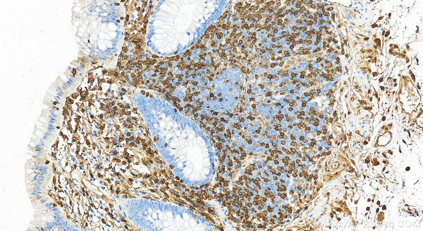
IHC staining of human colon using 21990-1-AP (same clone as 21990-1-PBS)
Immunohistochemical analysis of paraffin-embedded human normal colon slide using 21990-1-AP (Annexin A1 antibody) at dilution of 1:5000 (under 20x lens). Heat mediated antigen retrieval with Tris-EDTA buffer (pH 9.0). This data was developed using the same antibody clone with 21990-1-PBS in a different storage buffer formulation.
IHC staining of human colon using 21990-1-AP (same clone as 21990-1-PBS)
Immunohistochemical analysis of paraffin-embedded human normal colon slide using 21990-1-AP (Annexin A1 antibody) at dilution of 1:5000 (under 20x lens). Heat mediated antigen retrieval with Tris-EDTA buffer (pH 9.0). This data was developed using the same antibody clone with 21990-1-PBS in a different storage buffer formulation.
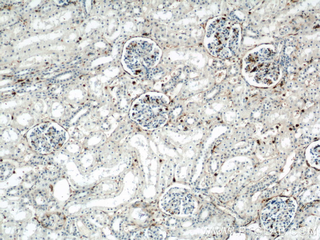
IHC staining of human kidney using 21990-1-AP (same clone as 21990-1-PBS)
Immunohistochemical analysis of paraffin-embedded human kidney using 21990-1-AP (Annexin A1 antibody) at dilution of 1:100 (under 10x lens). This data was developed using the same antibody clone with 21990-1-PBS in a different storage buffer formulation.
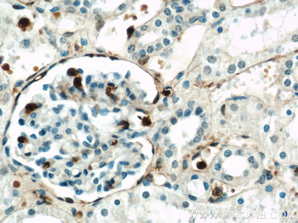
IHC staining of human kidney using 21990-1-AP (same clone as 21990-1-PBS)
Immunohistochemical analysis of paraffin-embedded human kidney using 21990-1-AP (Annexin A1 antibody) at dilution of 1:100 (under 40x lens). This data was developed using the same antibody clone with 21990-1-PBS in a different storage buffer formulation.
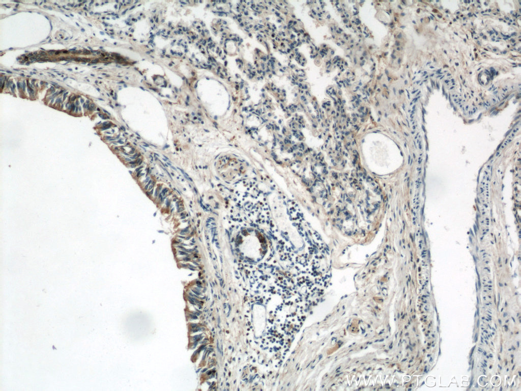
IHC staining of human lung using 21990-1-AP (same clone as 21990-1-PBS)
Immunohistochemical analysis of paraffin-embedded human lung using 21990-1-AP (Annexin A1 antibody) at dilution of 1:100 (under 10x lens). This data was developed using the same antibody clone with 21990-1-PBS in a different storage buffer formulation.
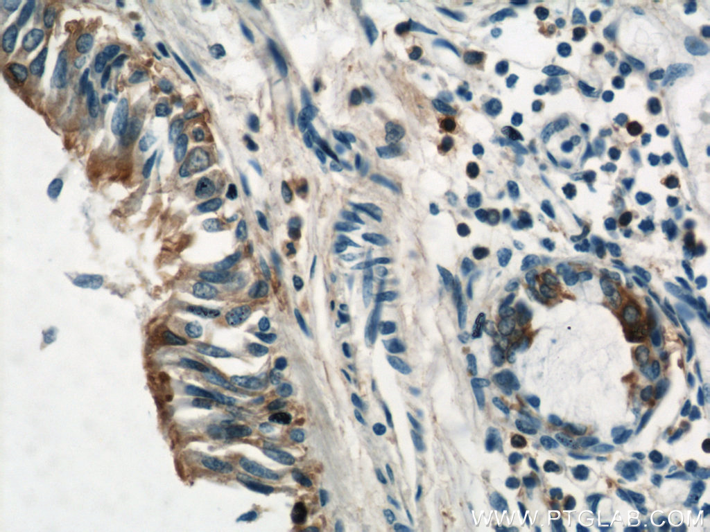
IHC staining of human lung using 21990-1-AP (same clone as 21990-1-PBS)
Immunohistochemical analysis of paraffin-embedded human lung using 21990-1-AP (Annexin A1 antibody) at dilution of 1:100 (under 40x lens). This data was developed using the same antibody clone with 21990-1-PBS in a different storage buffer formulation.
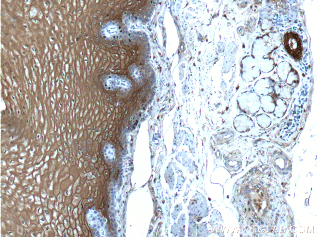
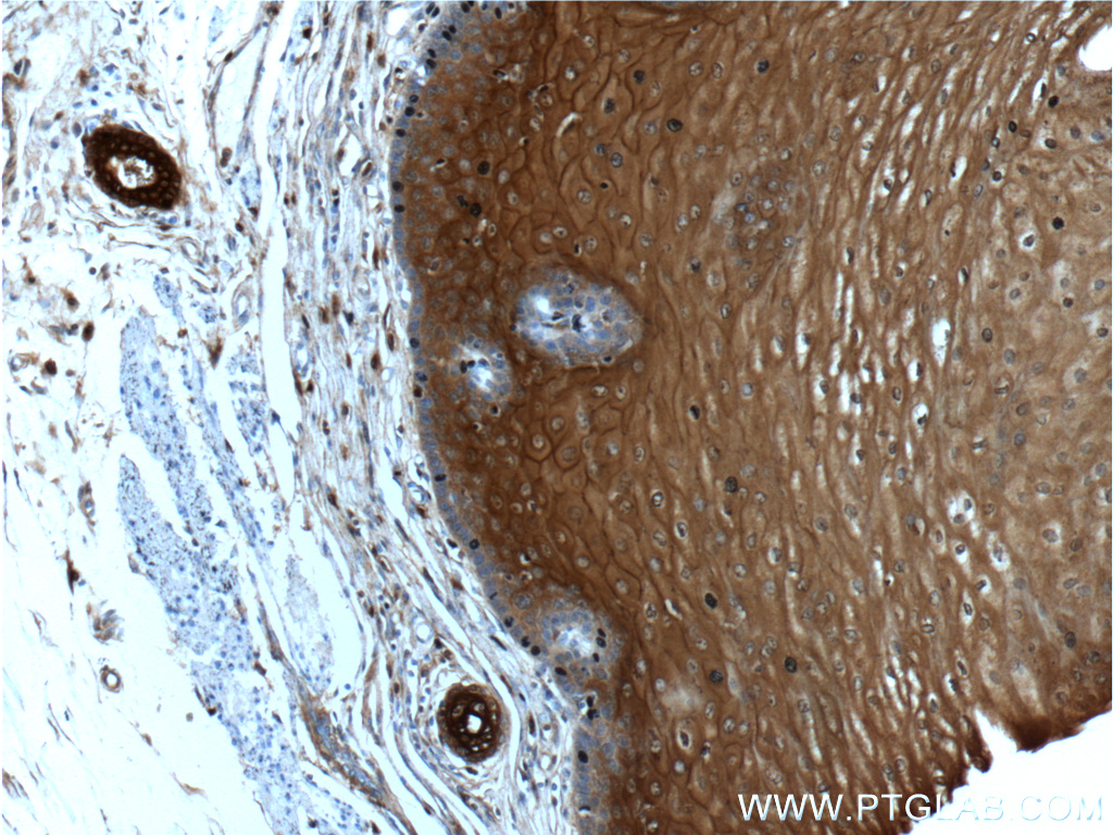
IHC staining of human oesophagus using 21990-1-AP (same clone as 21990-1-PBS)
Immunohistochemical analysis of paraffin-embedded human oesophagus tissue slide using 21990-1-AP (Annexin A1 antibody at dilution of 1:200 (under 10x lens). This data was developed using the same antibody clone with 21990-1-PBS in a different storage buffer formulation.
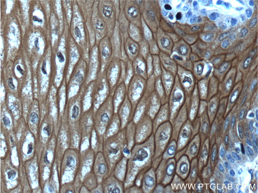
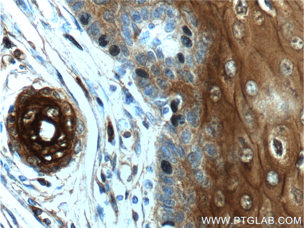
IHC staining of human oesophagus using 21990-1-AP (same clone as 21990-1-PBS)
Immunohistochemical analysis of paraffin-embedded human oesophagus tissue slide using 21990-1-AP (Annexin A1 antibody at dilution of 1:200 (under 40x lens). This data was developed using the same antibody clone with 21990-1-PBS in a different storage buffer formulation.
IHC staining of human oesophagus using 21990-1-AP (same clone as 21990-1-PBS)
Immunohistochemical analysis of paraffin-embedded human oesophagus tissue slide using 21990-1-AP (Annexin A1 antibody at dilution of 1:200 (under 10x lens). This data was developed using the same antibody clone with 21990-1-PBS in a different storage buffer formulation.
IHC staining of human oesophagus using 21990-1-AP (same clone as 21990-1-PBS)
Immunohistochemical analysis of paraffin-embedded human oesophagus tissue slide using 21990-1-AP (Annexin A1 antibody at dilution of 1:200 (under 40x lens). This data was developed using the same antibody clone with 21990-1-PBS in a different storage buffer formulation.
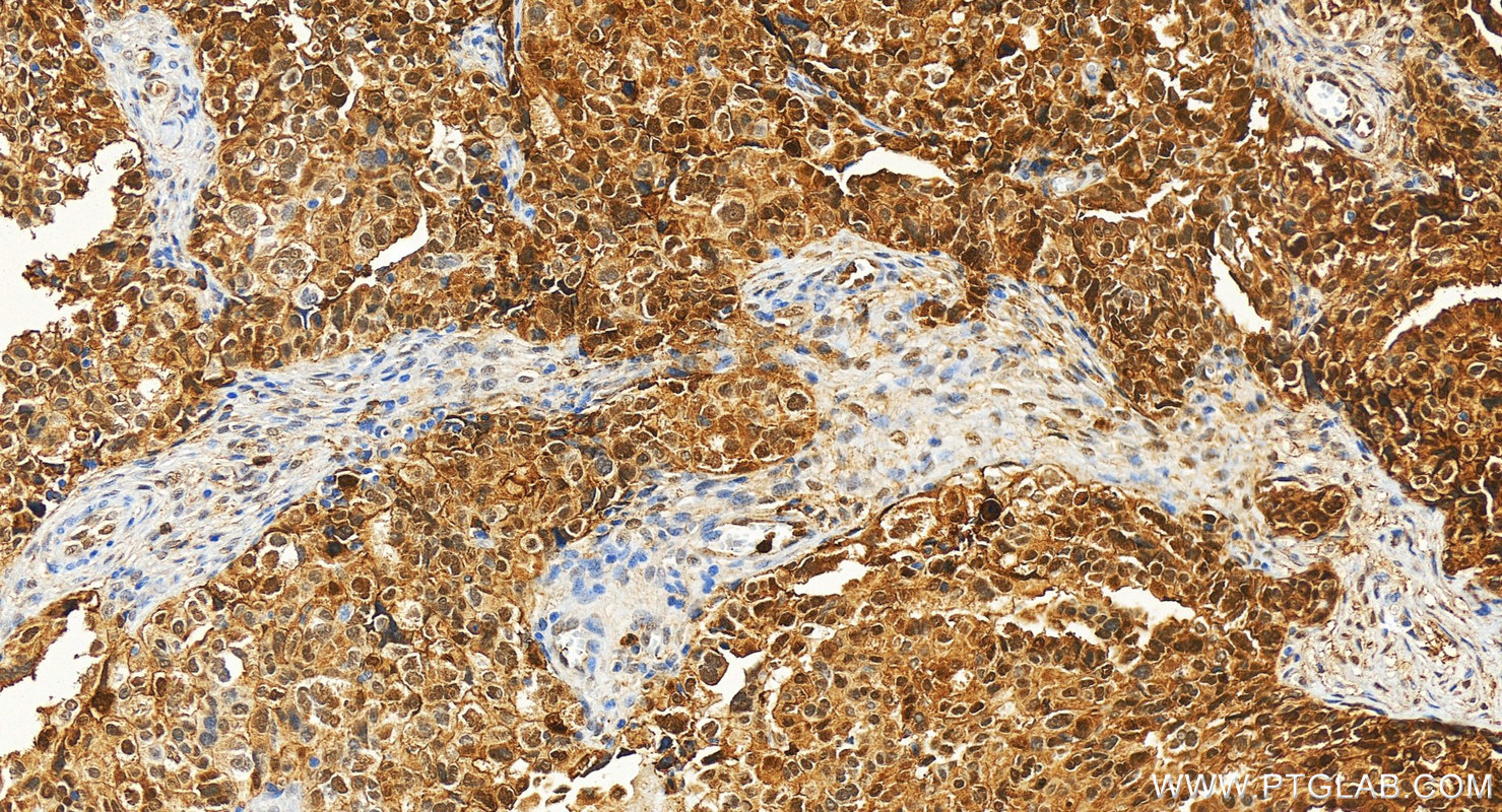
IHC staining of human ovary cancer using 21990-1-AP (same clone as 21990-1-PBS)
Immunohistochemical analysis of paraffin-embedded human ovary cancer tissue slide using 21990-1-AP (Annexin A1 antibody) at dilution of 1:5000 (under 20x lens). Heat mediated antigen retrieval with Tris-EDTA buffer (pH 9.0). This data was developed using the same antibody clone with 21990-1-PBS in a different storage buffer formulation.
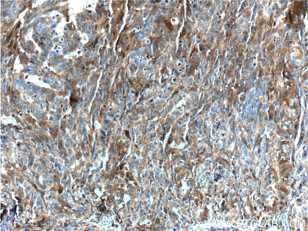
IHC staining of human ovary tumor using 21990-1-AP (same clone as 21990-1-PBS)
Immunohistochemical analysis of paraffin-embedded human ovary tumor tissue slide using 21990-1-AP (Annexin A1 antibody at dilution of 1:200 (under 10x lens). This data was developed using the same antibody clone with 21990-1-PBS in a different storage buffer formulation.
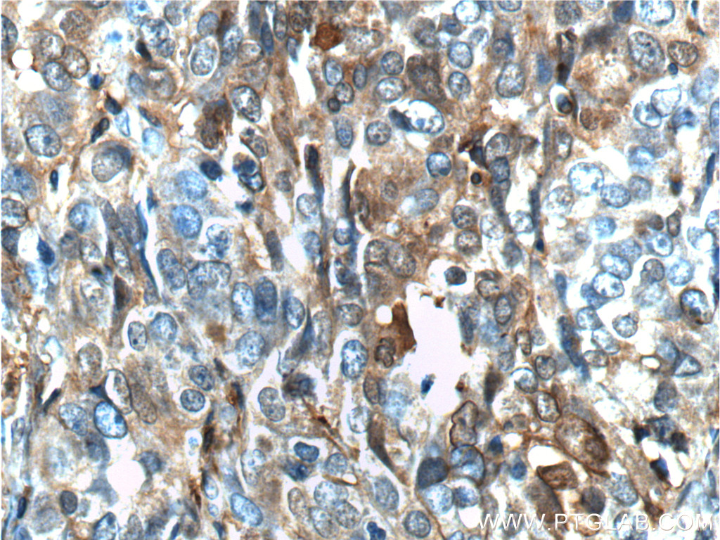
IHC staining of human ovary tumor using 21990-1-AP (same clone as 21990-1-PBS)
Immunohistochemical analysis of paraffin-embedded human ovary tumor tissue slide using 21990-1-AP (Annexin A1 antibody at dilution of 1:200 (under 40x lens). This data was developed using the same antibody clone with 21990-1-PBS in a different storage buffer formulation.
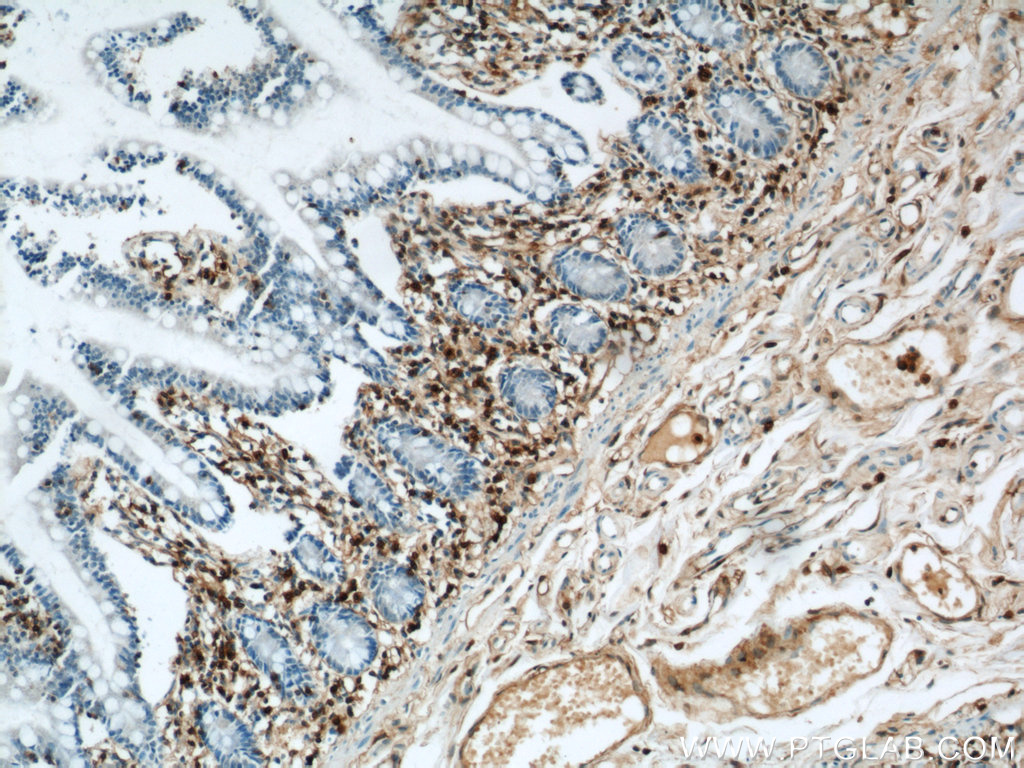
IHC staining of human small intestine using 21990-1-AP (same clone as 21990-1-PBS)
Immunohistochemical analysis of paraffin-embedded human small intestine using 21990-1-AP (Annexin A1 antibody) at dilution of 1:100 (under 10x lens). This data was developed using the same antibody clone with 21990-1-PBS in a different storage buffer formulation.
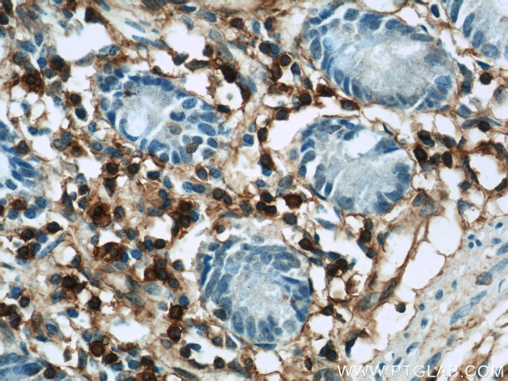
IHC staining of human small intestine using 21990-1-AP (same clone as 21990-1-PBS)
Immunohistochemical analysis of paraffin-embedded human small intestine using 21990-1-AP (Annexin A1 antibody) at dilution of 1:100 (under 40x lens). This data was developed using the same antibody clone with 21990-1-PBS in a different storage buffer formulation.
IF/ICC Figures
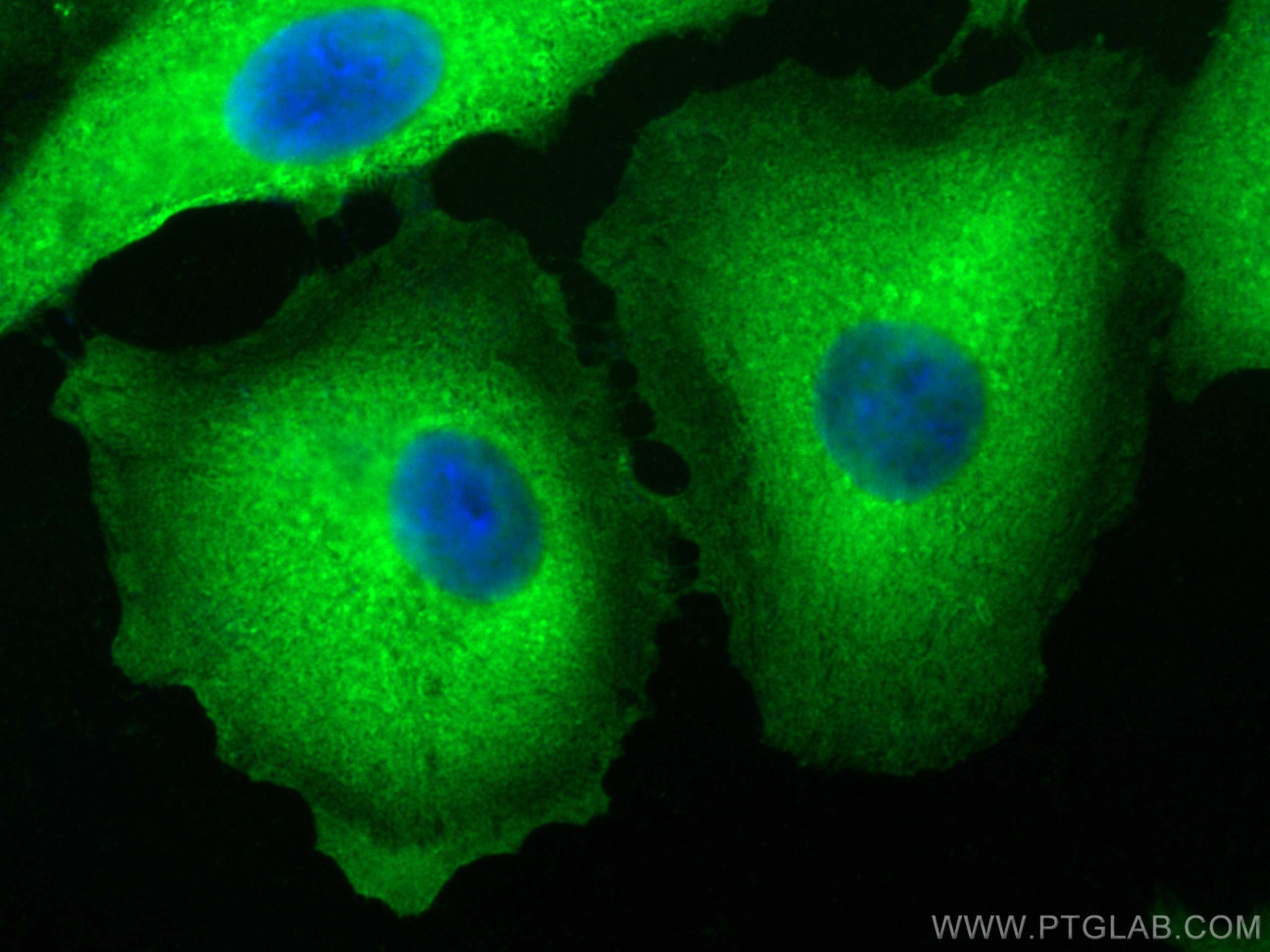
IF Staining of A431 using 21990-1-AP (same clone as 21990-1-PBS)
Immunofluorescent analysis of (-20°C Methanol) fixed A431 cells using Annexin A1 antibody (21990-1-AP) at dilution of 1:1000 and CoraLite®488-Conjugated Goat Anti-Rabbit IgG(H+L) (SA00013-2). This data was developed using the same antibody clone with 21990-1-PBS in a different storage buffer formulation.
IF Staining of A549 using 21990-1-AP (same clone as 21990-1-PBS)
Immunofluorescent analysis of (-20°C Methanol) fixed A549 cells using Annexin A1 antibody (21990-1-AP) at dilution of 1:400 and CoraLite®488-Conjugated AffiniPure Goat Anti-Rabbit IgG(H+L) (SA00013-2). This data was developed using the same antibody clone with 21990-1-PBS in a different storage buffer formulation.
FC (INTRA) Figures
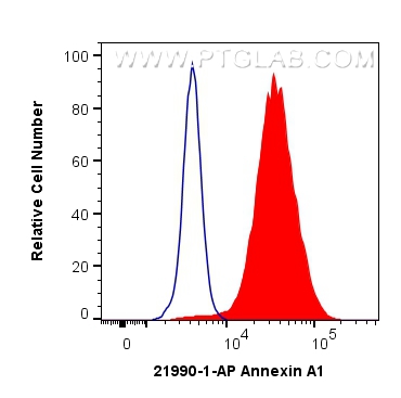
FC experiment of Jurkat using 21990-1-AP (same clone as 21990-1-PBS)
1x10^6 Jurkat cells were intracellularly stained with 0.25 ug Annexin A1 Polyclonal antibody (21990-1-AP) and CoraLite®488-Conjugated Goat Anti-Rabbit IgG(H+L) (SA00013-2)(red), or 0.25 ug rabbit IgG isotype control (blue). Cells were fixed and permeabilized with Transcription Factor Staining Buffer Kit (PF00011). This data was developed using the same antibody clone with 21990-1-PBS in a different storage buffer formulation.
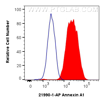
FC experiment of NIH/3T3 using 21990-1-AP (same clone as 21990-1-PBS)
1X10^6 NIH/3T3 cells were intracellularly stained with 0.5 ug Anti-Human Annexin A1 (21990-1-AP) and CoraLite®488-Conjugated AffiniPure Goat Anti-Rabbit IgG(H+L) at dilution 1:1000 (red), or 0.5 ug Control Antibody. Cells were fixed with 4% PFA and permeabilized with Flow Cytometry Perm Buffer. This data was developed using the same antibody clone with 21990-1-PBS in a different storage buffer formulation.