WB Figures
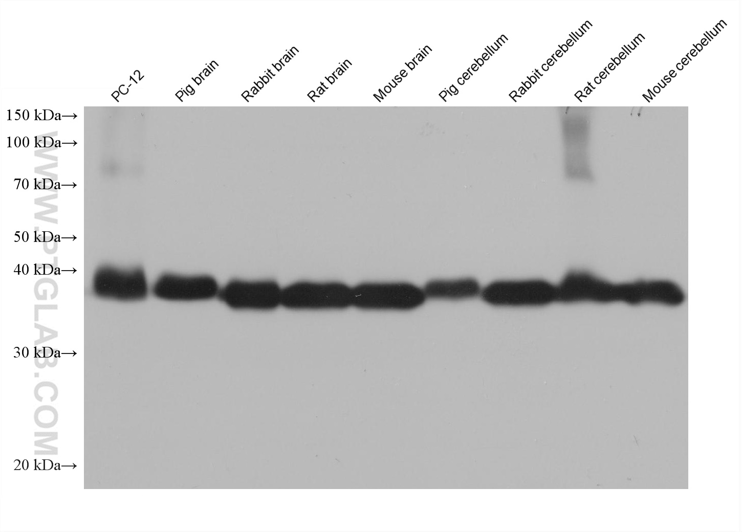
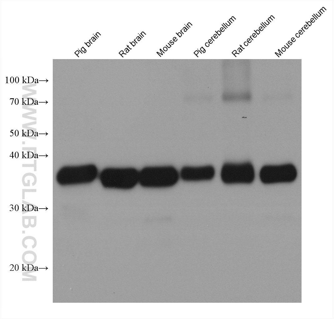
WB analysis using 67864-1-Ig
Various lysates were subjected to SDS PAGE followed by western blot with 67864-1-Ig (Synaptophysin antibody) at dilution of 1:100000 incubated at room temperature for 1.5 hours.
WB analysis using 67864-1-Ig
Various lysates were subjected to SDS PAGE followed by western blot with 67864-1-Ig (Synaptophysin antibody) at dilution of 1:100000 incubated at room temperature for 1.5 hours.
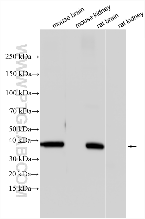
WB analysis using 67864-1-Ig
Various lysates were subjected to SDS PAGE followed by western blot with 67864-1-Ig (Synaptophysin antibody) at dilution of 1:50000 incubated at room temperature for 1.5 hours.
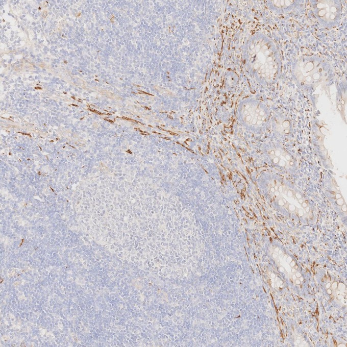
IHC staining of human appendicitis using 67864-1-Ig
Immunohistochemical analysis of paraffin-embedded human appendicitis tissue slide using 67864-1-Ig (Synaptophysin antibody) at dilution of 1:5000. Heat mediated antigen retrieval with Tris-EDTA buffer (pH 9.0).
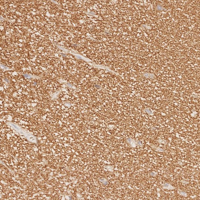
IHC staining of human brain using 67864-1-Ig
Immunohistochemical analysis of paraffin-embedded human brain tissue slide using 67864-1-Ig (Synaptophysin antibody) at dilution of 1:5000. Heat mediated antigen retrieval with Tris-EDTA buffer (pH 9.0).
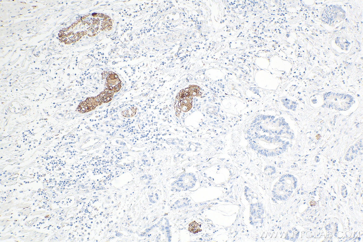
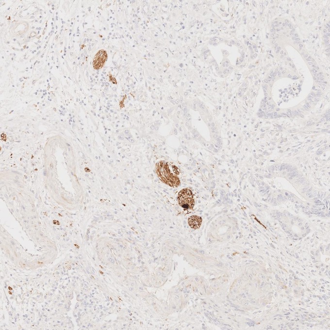
IHC staining of human colorectal cancer using 67864-1-Ig
Immunohistochemical analysis of paraffin-embedded human colorectal cancer tissue slide using 67864-1-Ig (Synaptophysin antibody) at dilution of 1:5000. Heat mediated antigen retrieval with Tris-EDTA buffer (pH 9.0).
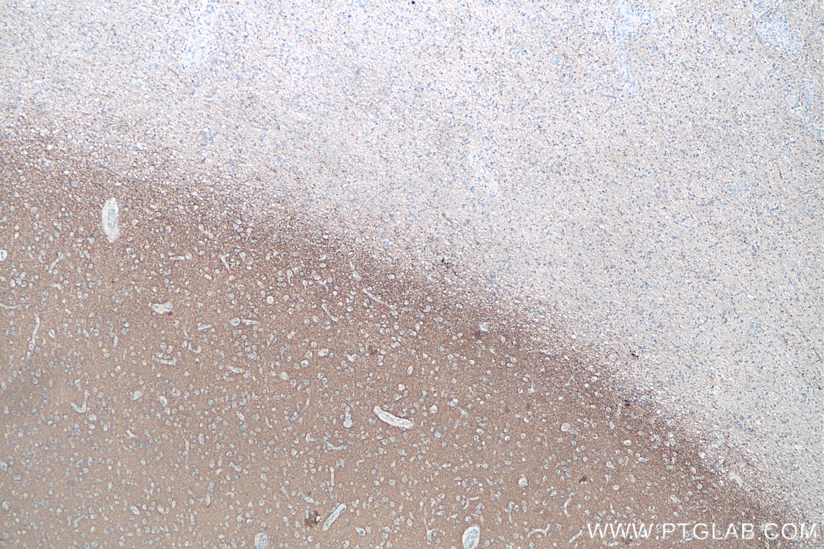
IHC staining of human gliomas using 67864-1-Ig
Immunohistochemical analysis of paraffin-embedded human gliomas tissue slide using 67864-1-Ig (Synaptophysin antibody) at dilution of 1:4000 (under 4x lens). Heat mediated antigen retrieval with Tris-EDTA buffer (pH 9.0).
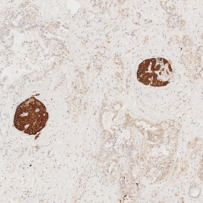
IHC staining of human pancreas cancer using 67864-1-Ig
Immunohistochemical analysis of paraffin-embedded human pancreas cancer tissue slide using 67864-1-Ig (Synaptophysin antibody) at dilution of 1:5000. Heat mediated antigen retrieval with Tris-EDTA buffer (pH 9.0).
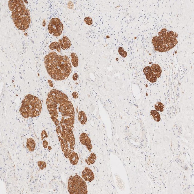
IHC staining of human pancreas using 67864-1-Ig
Immunohistochemical analysis of paraffin-embedded human pancreas tissue slide using 67864-1-Ig (Synaptophysin antibody) at dilution of 1:5000. Heat mediated antigen retrieval with Tris-EDTA buffer (pH 9.0).
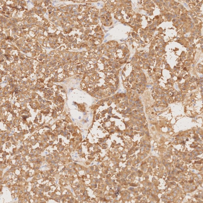
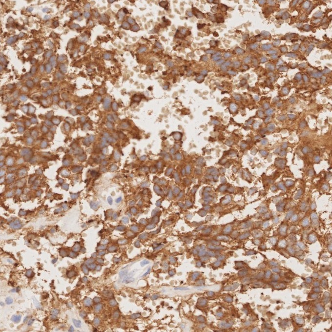
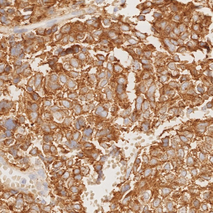
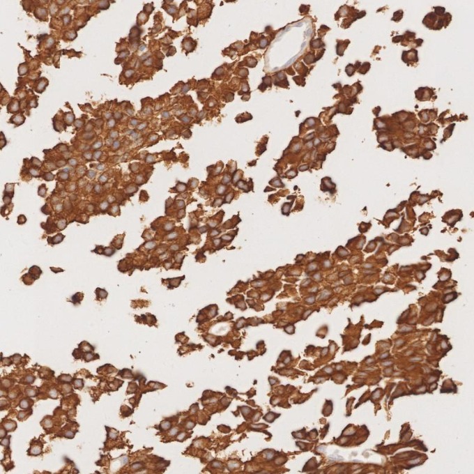
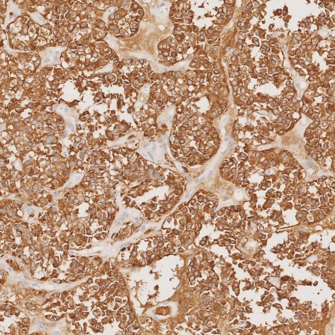
IHC staining of human pituitary adenoma using 67864-1-Ig
Immunohistochemical analysis of paraffin-embedded human pituitary adenoma tissue slide using 67864-1-Ig (Synaptophysin antibody) at dilution of 1:5000. Heat mediated antigen retrieval with Tris-EDTA buffer (pH 9.0).
IHC staining of human pituitary adenoma using 67864-1-Ig
Immunohistochemical analysis of paraffin-embedded human pituitary adenoma tissue slide using 67864-1-Ig (Synaptophysin antibody) at dilution of 1:5000. Heat mediated antigen retrieval with Tris-EDTA buffer (pH 9.0).
IHC staining of human pituitary adenoma using 67864-1-Ig
Immunohistochemical analysis of paraffin-embedded human pituitary adenoma tissue slide using 67864-1-Ig (Synaptophysin antibody) at dilution of 1:5000. Heat mediated antigen retrieval with Tris-EDTA buffer (pH 9.0).
IHC staining of human rectal cancer using 67864-1-Ig
Immunohistochemical analysis of paraffin-embedded human rectal cancer tissue slide using 67864-1-Ig (Synaptophysin antibody) at dilution of 1:4000 (under 10x lens). Heat mediated antigen retrieval with Tris-EDTA buffer (pH 9.0).
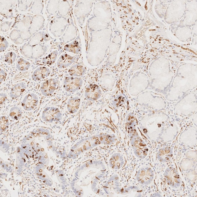
IHC staining of human stomach cancer using 67864-1-Ig
Immunohistochemical analysis of paraffin-embedded human stomach cancer tissue slide using 67864-1-Ig (Synaptophysin antibody) at dilution of 1:5000. Heat mediated antigen retrieval with Tris-EDTA buffer (pH 9.0).
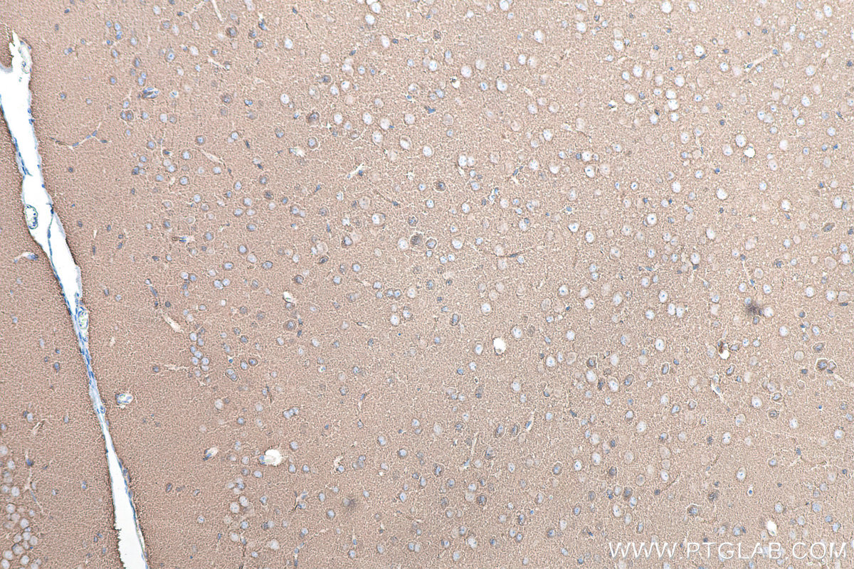
IHC staining of mouse brain using 67864-1-Ig
Immunohistochemical analysis of paraffin-embedded mouse brain tissue slide using 67864-1-Ig (Synaptophysin antibody) at dilution of 1:4000 (under 10x lens). Heat mediated antigen retrieval with Tris-EDTA buffer (pH 9.0).
IHC staining of mouse brain using 67864-1-Ig
Immunohistochemical analysis of paraffin-embedded mouse brain tissue slide using 67864-1-Ig (Synaptophysin antibody) at dilution of 1:1000 (under 40x lens). Heat mediated antigen retrieval with Tris-EDTA buffer (pH 9.0).
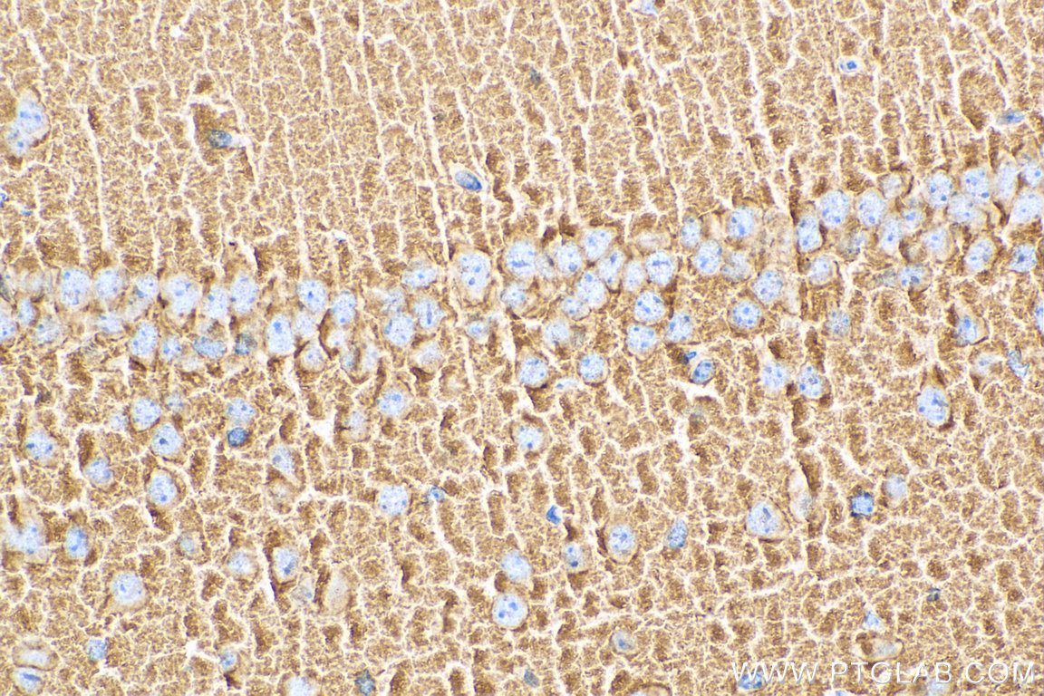
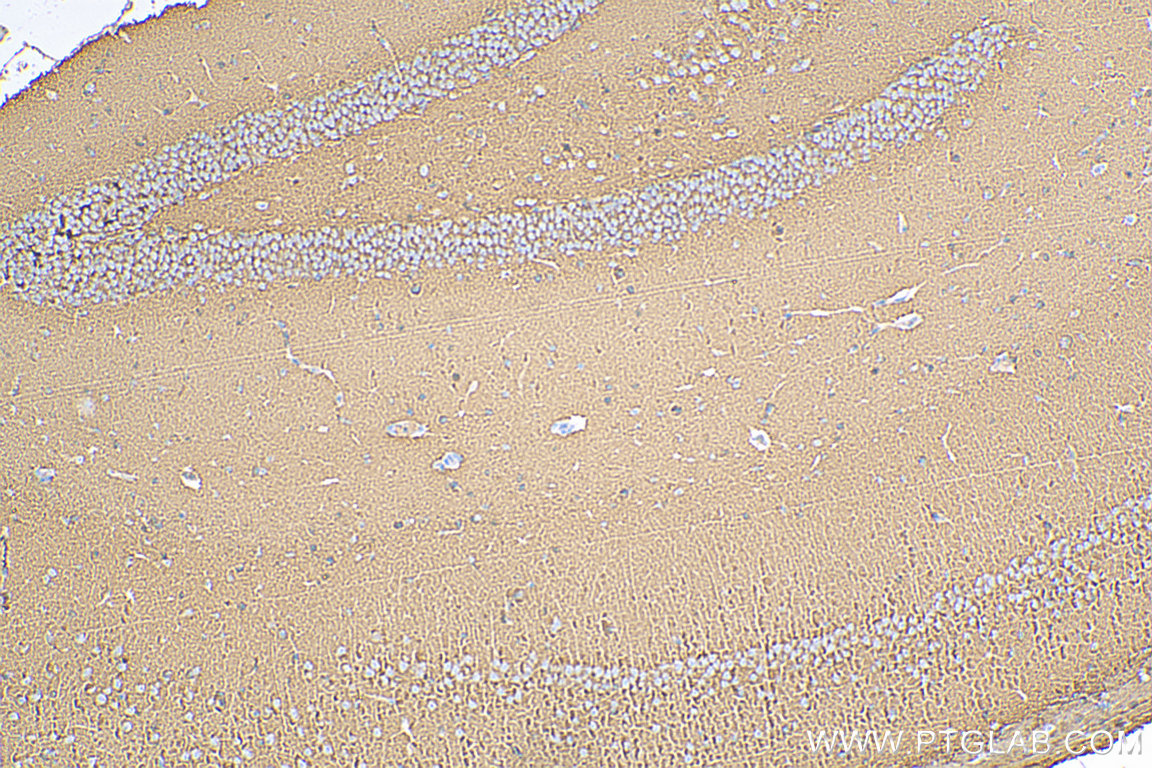
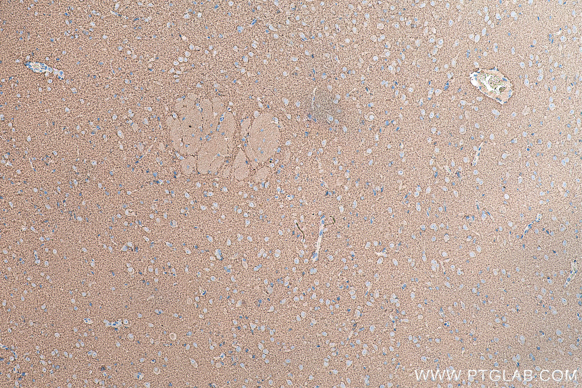
IHC staining of mouse brain using 67864-1-Ig
Immunohistochemical analysis of paraffin-embedded mouse brain tissue slide using 67864-1-Ig (Synaptophysin antibody) at dilution of 1:1000 (under 10x lens). Heat mediated antigen retrieval with Tris-EDTA buffer (pH 9.0).
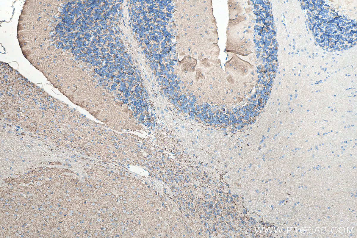
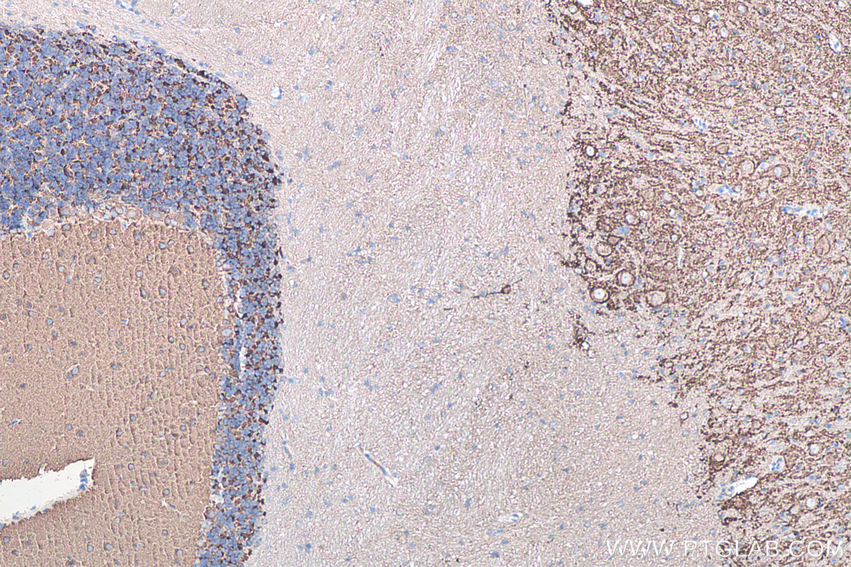
IHC staining of mouse cerebellum using 67864-1-Ig
Immunohistochemical analysis of paraffin-embedded mouse cerebellum tissue slide using 67864-1-Ig (Synaptophysin antibody) at dilution of 1:4000 (under 10x lens). Heat mediated antigen retrieval with Tris-EDTA buffer (pH 9.0).
IHC staining of rat brain using 67864-1-Ig
Immunohistochemical analysis of paraffin-embedded rat brain tissue slide using 67864-1-Ig (Synaptophysin antibody) at dilution of 1:4000 (under 10x lens). Heat mediated antigen retrieval with Tris-EDTA buffer (pH 9.0).
IHC staining of rat cerebellum using 67864-1-Ig
Immunohistochemical analysis of paraffin-embedded rat cerebellum tissue slide using 67864-1-Ig (Synaptophysin antibody) at dilution of 1:4000 (under 10x lens). Heat mediated antigen retrieval with Tris-EDTA buffer (pH 9.0).
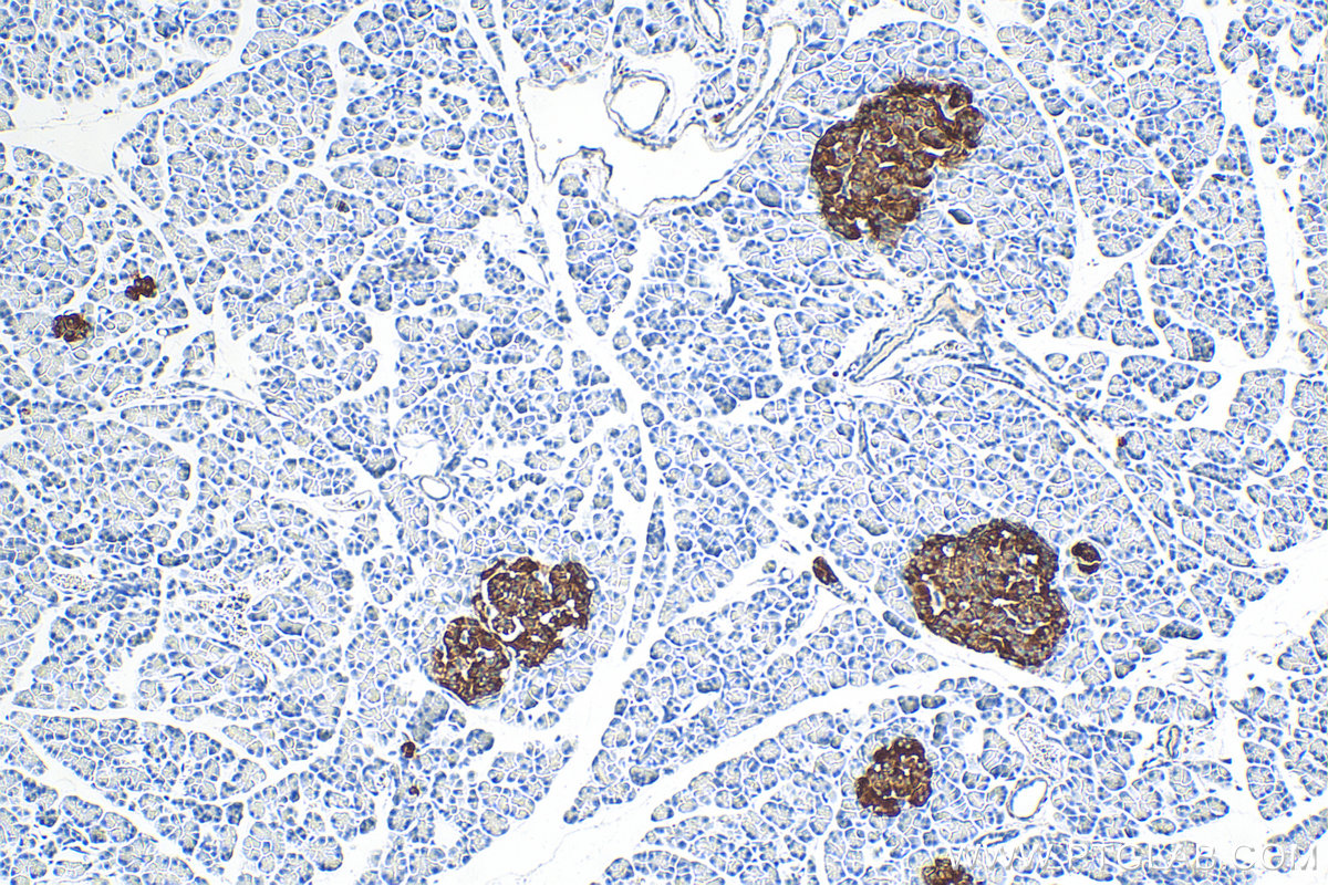
IHC staining of rat pancreas using 67864-1-Ig
Immunohistochemical analysis of paraffin-embedded rat pancreas tissue slide using 67864-1-Ig (Synaptophysin antibody) at dilution of 1:4000 (under 10x lens). Heat mediated antigen retrieval with Tris-EDTA buffer (pH 9.0).
IHC-AUTOSTAINER Figures
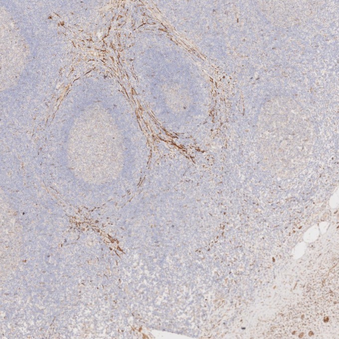
IHC staining of human appendicitis using 67864-1-Ig
Immunohistochemical analysis of paraffin-embedded human appendicitis tissue slide using 67864-1-Ig (Synaptophysin antibody) at dilution of 1:5000. Heat mediated antigen retrieval with Tris-EDTA buffer (pH 9.0). The staining was performed on an autostainer.
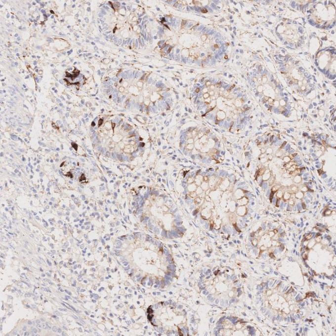
IHC staining of human colorectal cancer using 67864-1-Ig
Immunohistochemical analysis of paraffin-embedded human colorectal cancer tissue slide using 67864-1-Ig (Synaptophysin antibody) at dilution of 1:5000. Heat mediated antigen retrieval with Tris-EDTA buffer (pH 9.0). The staining was performed on an autostainer.
IHC staining of human pituitary adenoma using 67864-1-Ig
Immunohistochemical analysis of paraffin-embedded human pituitary adenoma tissue slide using 67864-1-Ig (Synaptophysin antibody) at dilution of 1:5000. Heat mediated antigen retrieval with Tris-EDTA buffer (pH 9.0). The staining was performed on an autostainer.
IHC staining of human pituitary adenoma using 67864-1-Ig
Immunohistochemical analysis of paraffin-embedded human pituitary adenoma tissue slide using 67864-1-Ig (Synaptophysin antibody) at dilution of 1:5000. Heat mediated antigen retrieval with Tris-EDTA buffer (pH 9.0). The staining was performed on an autostainer.
IF-P Figures
IF Staining of mouse brain using 67864-1-Ig
Immunofluorescent analysis of (4% PFA) fixed mouse brain tissue using Synaptophysin antibody (67864-1-Ig, Clone: 4E12C4 ) at dilution of 1:400 and CoraLite®488-Conjugated AffiniPure Goat Anti-Mouse IgG(H+L).
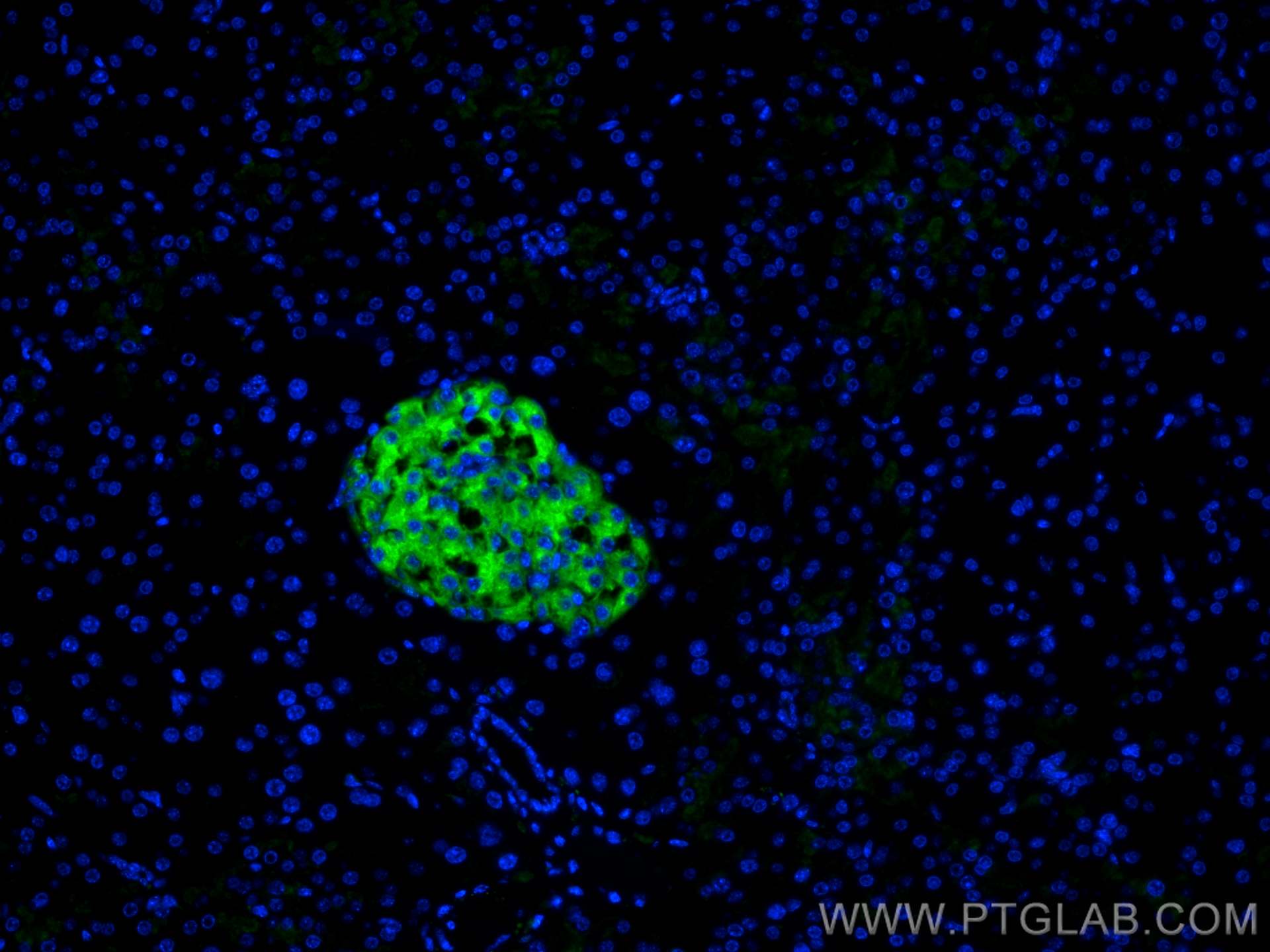
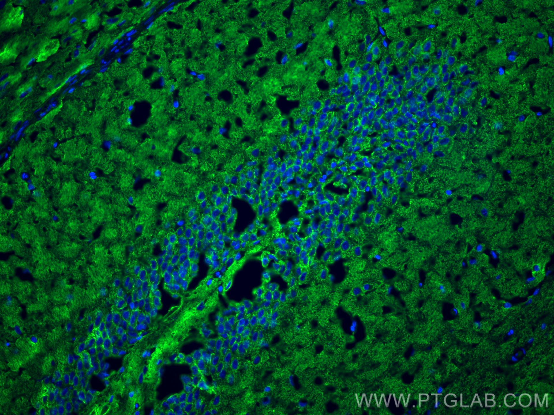
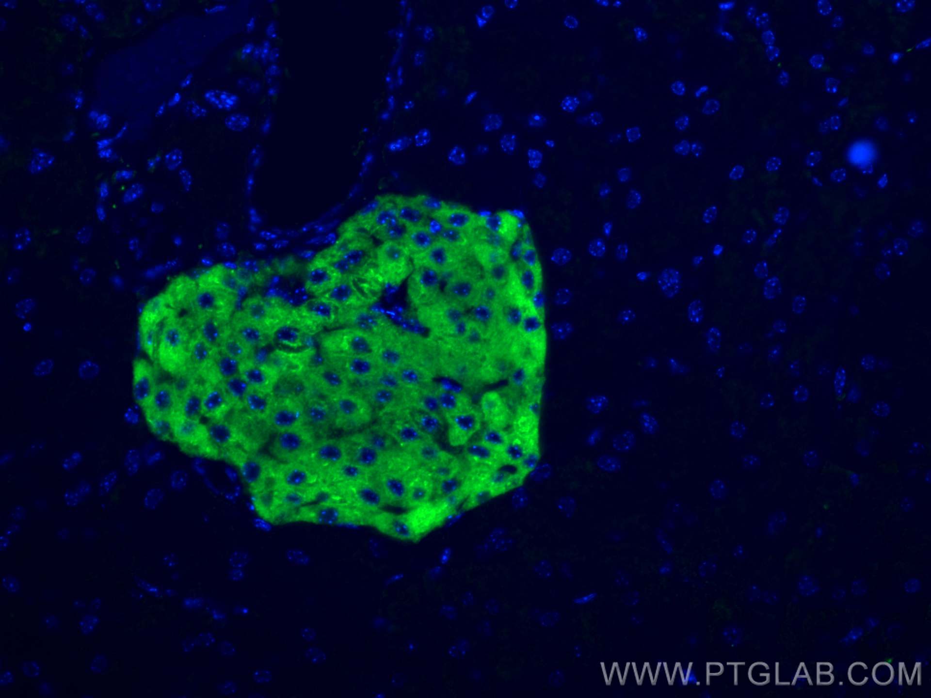
IF Staining of mouse pancreas using 67864-1-Ig
Immunofluorescent analysis of (4% PFA) fixed mouse pancreas tissue using Synaptophysin antibody (67864-1-Ig, Clone: 4E12C4 ) at dilution of 1:400 and CoraLite®488-Conjugated AffiniPure Goat Anti-Mouse IgG(H+L).
IF Staining of mouse pancreas using 67864-1-Ig
Immunofluorescent analysis of (4% PFA) fixed mouse pancreas tissue using Synaptophysin antibody (67864-1-Ig, Clone: 4E12C4 ) at dilution of 1:400 and CoraLite®488-Conjugated AffiniPure Goat Anti-Mouse IgG(H+L).
IF/ICC Figures
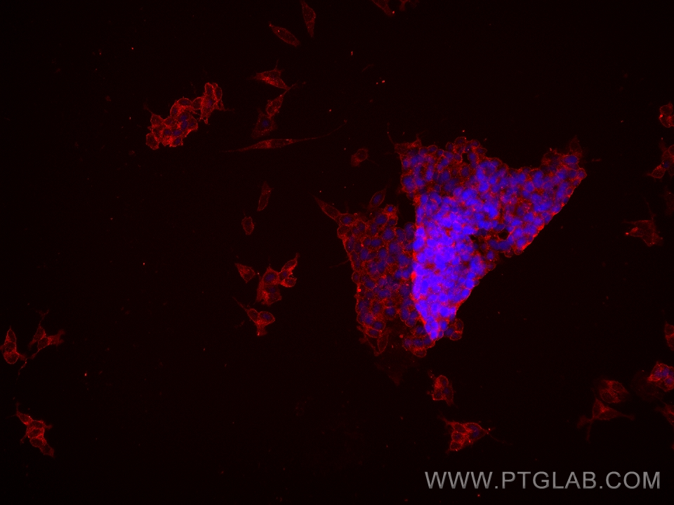
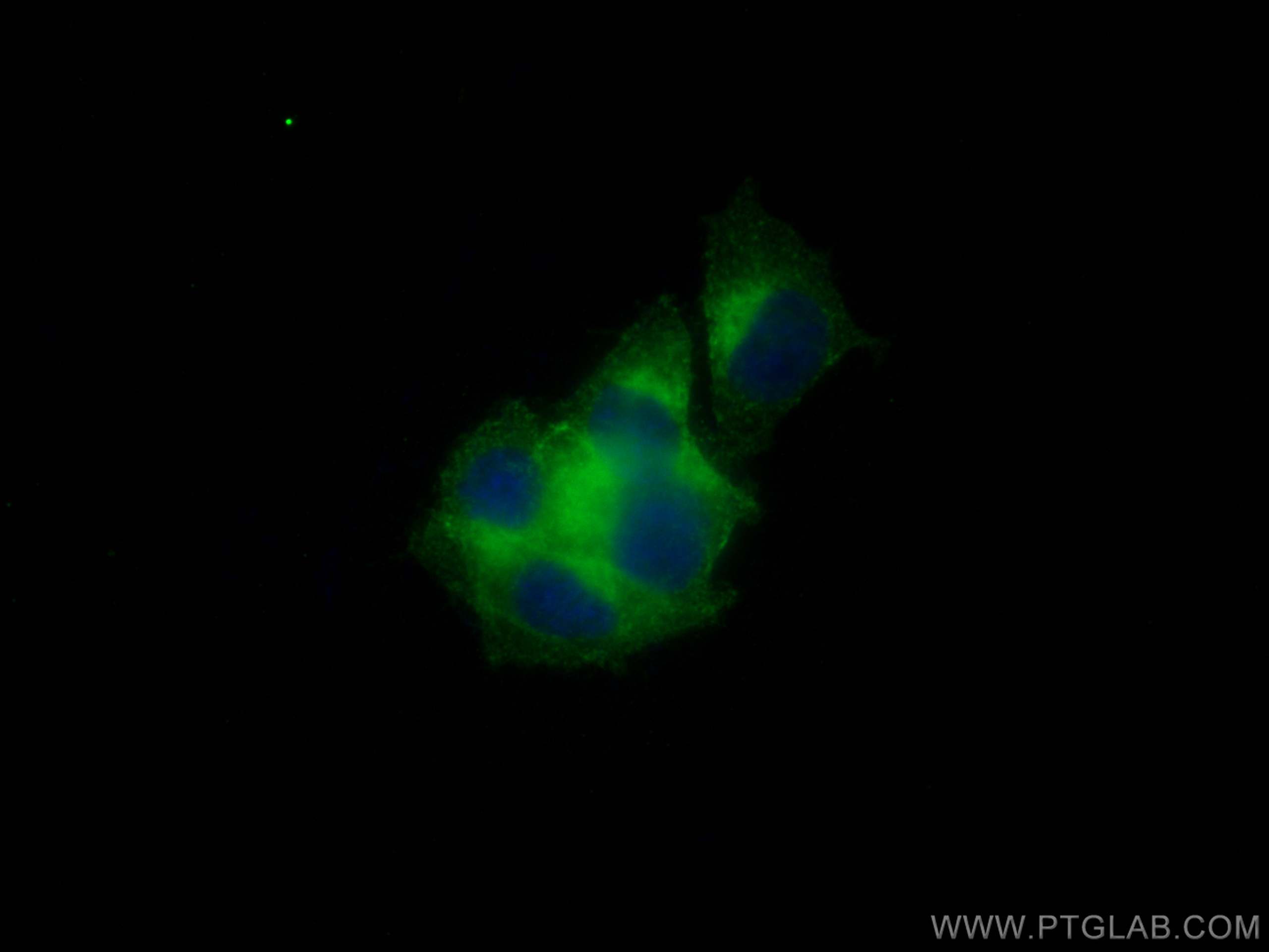
IF Staining of PC-12 using 67864-1-Ig
Immunofluorescent analysis of (4% PFA) fixed PC-12 cells using Synaptophysin antibody (67864-1-Ig, Clone: 4E12C4 ) at dilution of 1:800 and CoraLite®488-Conjugated AffiniPure Goat Anti-Mouse IgG(H+L).
IF Staining of SH-SY5Y using 67864-1-Ig
Immunofluorescent analysis of (4% PFA) fixed SH-SY5Y cells using Synaptophysin antibody (67864-1-Ig, Clone: 4E12C4 ) at dilution of 1:500 and Multi-rAb CoraLite® Plus 594-Goat Anti-Mouse Recombinant Secondary Antibody (H+L) (Cat.NO. RGAM004 ).