WB Figures
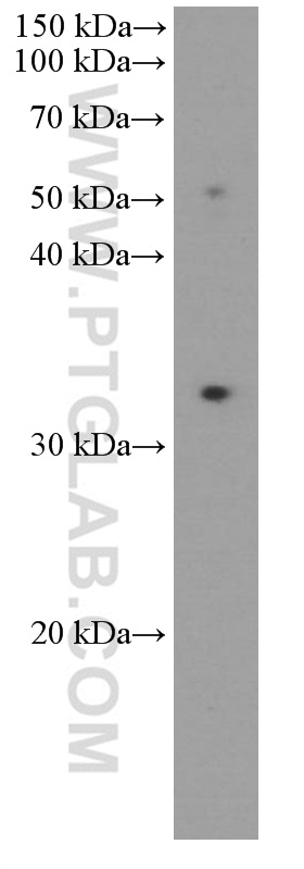
WB analysis of human lymph node using 66220-1-Ig
human lymph node tissue were subjected to SDS PAGE followed by western blot with 66220-1-Ig (PD-1/CD279 antibody) at dilution of 1:1000 incubated at room temperature for 1.5 hours.
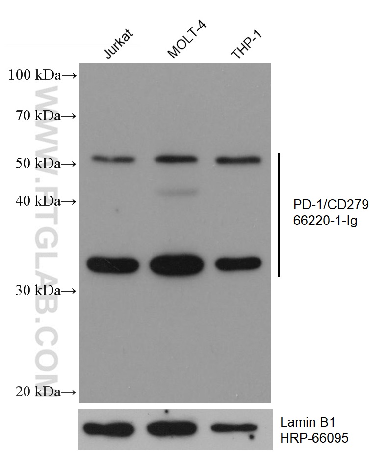
WB analysis using 66220-1-Ig
Various lysates were subjected to SDS PAGE followed by western blot with 66220-1-Ig (PD-1/CD279 antibody) at dilution of 1:15000 incubated at room temperature for 1.5 hours. The membrane was stripped and reblotted with HRP-conjugated Lamin B1 Monoclonal antibody (HRP-66095) as loading control.
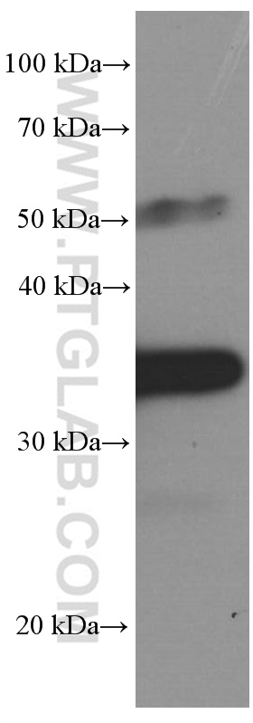
WB analysis of mouse thymus using 66220-1-Ig
mouse thymus tissue was subjected to SDS PAGE followed by western blot with 66220-1-Ig (PD-1/CD279 Antibody) at dilution of 1:5000 incubated at room temperature for 1.5 hours.
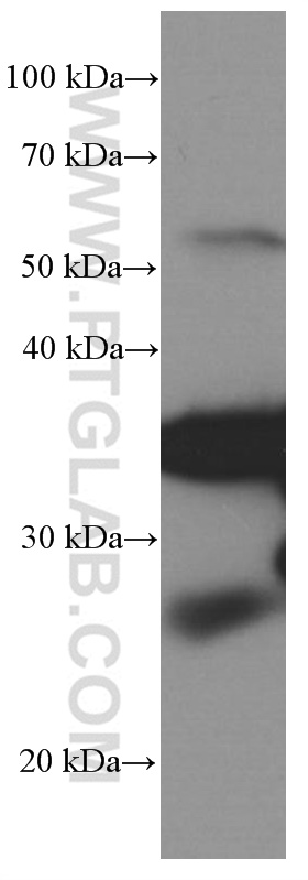
WB analysis of rat spleen using 66220-1-Ig
rat spleen tissue were subjected to SDS PAGE followed by western blot with 66220-1-Ig (PD-1/CD279 Antibody) at dilution of 1:5000 incubated at room temperature for 1.5 hours.
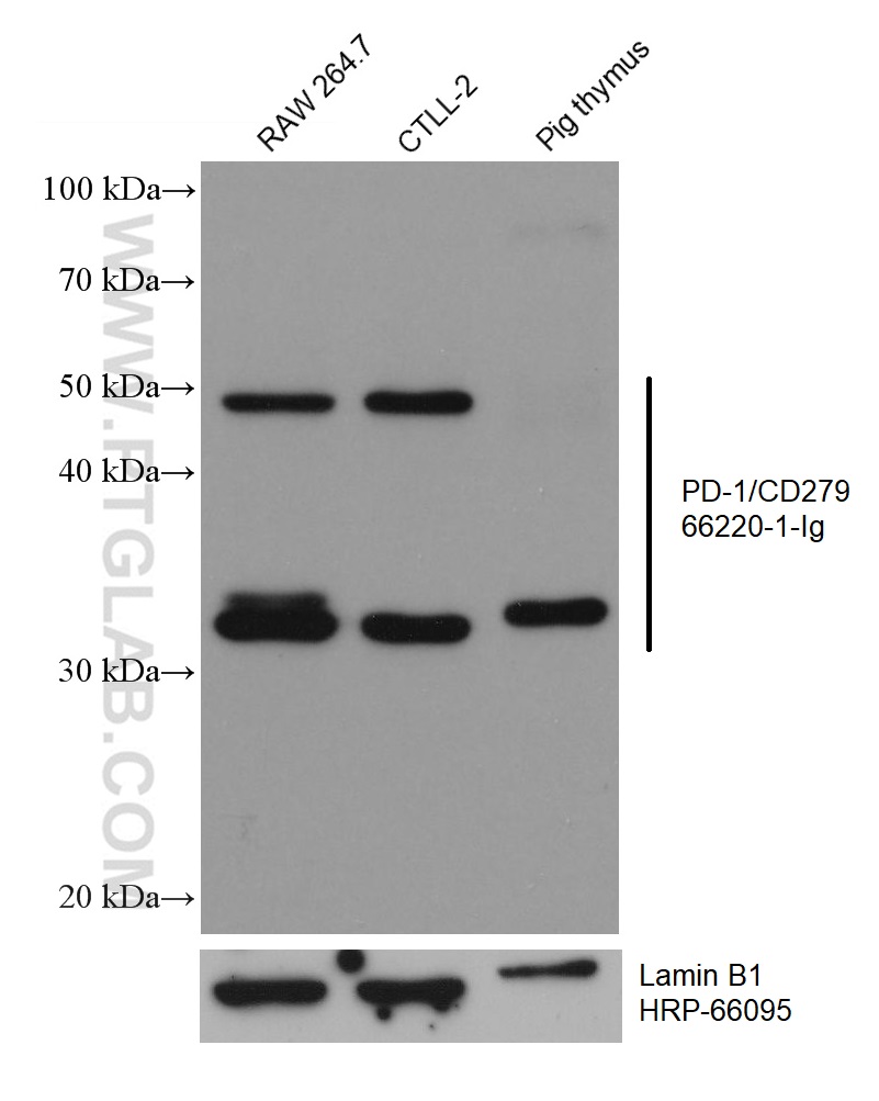
WB analysis using 66220-1-Ig
Various lysates were subjected to SDS PAGE followed by western blot with 66220-1-Ig (PD-1/CD279 antibody) at dilution of 1:15000 incubated at room temperature for 1.5 hours. The membrane was stripped and reblotted with HRP-conjugated Lamin B1 Monoclonal antibody (HRP-66095) as loading control.
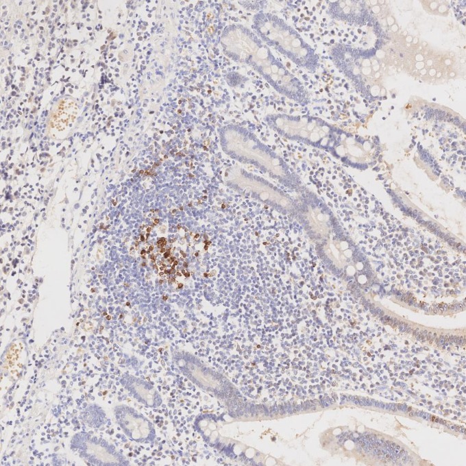
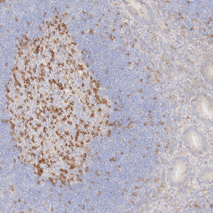
IHC staining of human appendicitis using 66220-1-Ig
Immunohistochemical analysis of paraffin-embedded human appendicitis tissue slide using 66220-1-Ig (PD-1/CD279 antibody) at dilution of 1:20000. Heat mediated antigen retrieval with Tris-EDTA buffer (pH 9.0).
IHC staining of human appendicitis using 66220-1-Ig
Immunohistochemical analysis of paraffin-embedded human appendicitis tissue slide using 66220-1-Ig (PD-1/CD279 antibody) at dilution of 1:20000. Heat mediated antigen retrieval with Tris-EDTA buffer (pH 9.0).
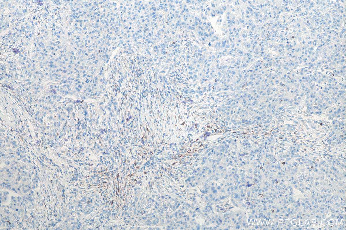
IHC staining of human breast cancer using 66220-1-Ig
Immunohistochemical analysis of paraffin-embedded human breast cancer tissue slide using 66220-1-Ig (PD-1/CD279 antibody) at dilution of 1:10000 (under 10x lens). Heat mediated antigen retrieval with Tris-EDTA buffer (pH 9.0).
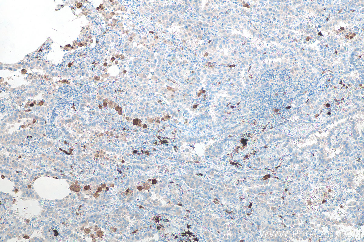
IHC staining of human lung cancer using 66220-1-Ig
Immunohistochemical analysis of paraffin-embedded human lung cancer tissue slide using 66220-1-Ig (PD-1/CD279 antibody) at dilution of 1:10000 (under 10x lens). Heat mediated antigen retrieval with Tris-EDTA buffer (pH 9.0).
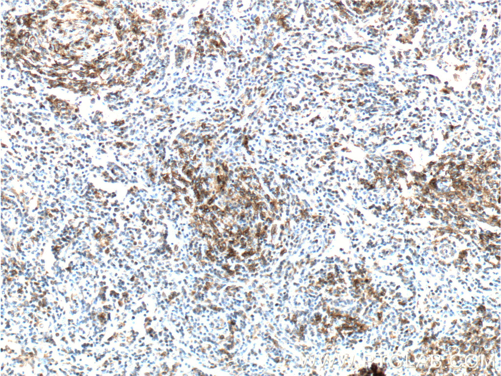
IHC staining of human lymphoma using 66220-1-Ig
Immunohistochemical analysis of paraffin-embedded human angioimmunoblastic T-cell lymphoma tissue slide using 66220-1-Ig (PD-1/CD279 Antibody) at dilution of 1:200 (under 10x lens). . Heat mediated antigen retrieved with Tris-EDTA buffer, pH9.0.
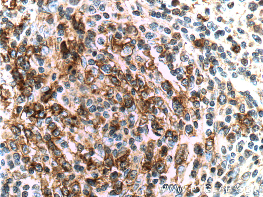
IHC staining of human lymphoma using 66220-1-Ig
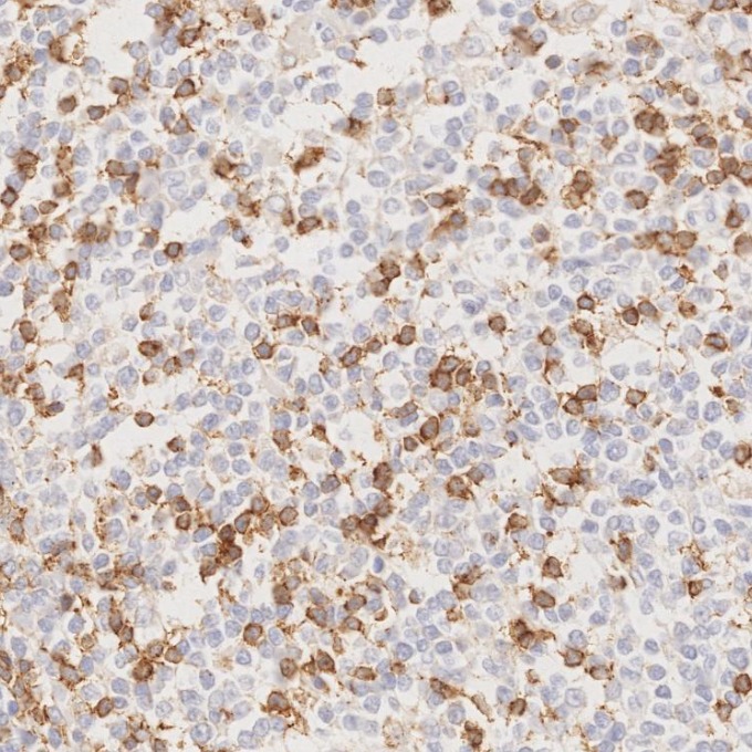
Immunohistochemical analysis of paraffin-embedded human angioimmunoblastic T-cell lymphoma tissue slide using 66220-1-Ig (PD-1/CD279 Antibody) at dilution of 1:200 (under 40x lens). . Heat mediated antigen retrieved with Tris-EDTA buffer, pH9.0.
IHC staining of human lymphoma using 66220-1-Ig
Immunohistochemical analysis of paraffin-embedded human lymphoma tissue slide using 66220-1-Ig (PD-1/CD279 antibody) at dilution of 1:20000. Heat mediated antigen retrieval with Tris-EDTA buffer (pH 9.0).
IHC staining of human lymphoma using 66220-1-Ig
Immunohistochemical analysis of paraffin-embedded human lymphoma tissue slide using 66220-1-Ig (PD-1/CD279 antibody) at dilution of 1:20000. Heat mediated antigen retrieval with Tris-EDTA buffer (pH 9.0).
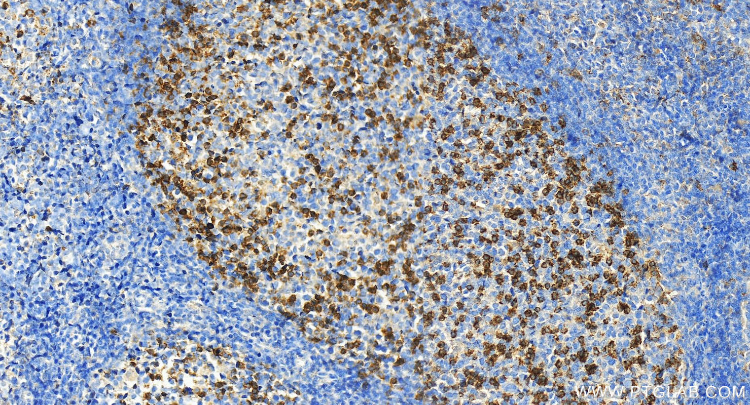
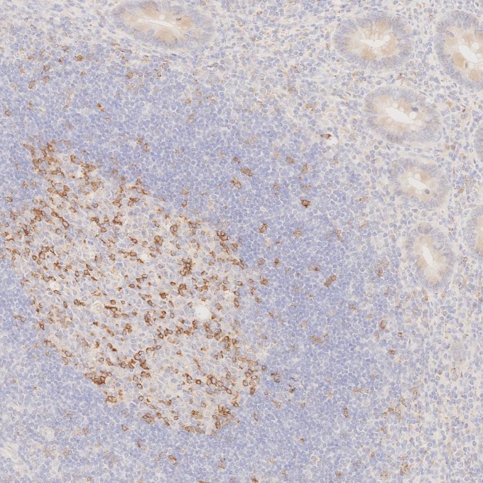
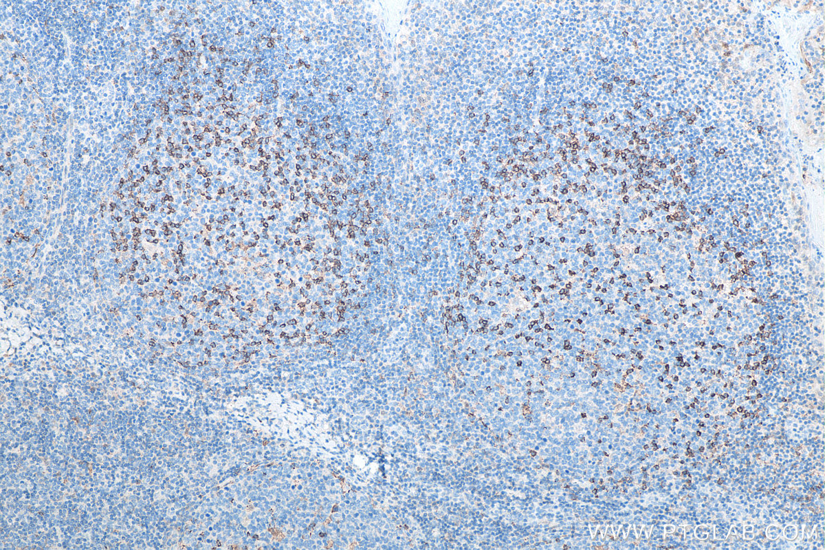
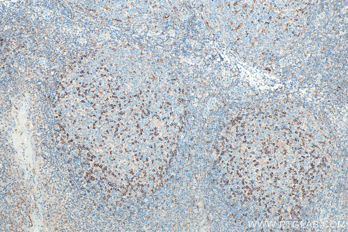
IHC staining of human tonsillitis using 66220-1-Ig
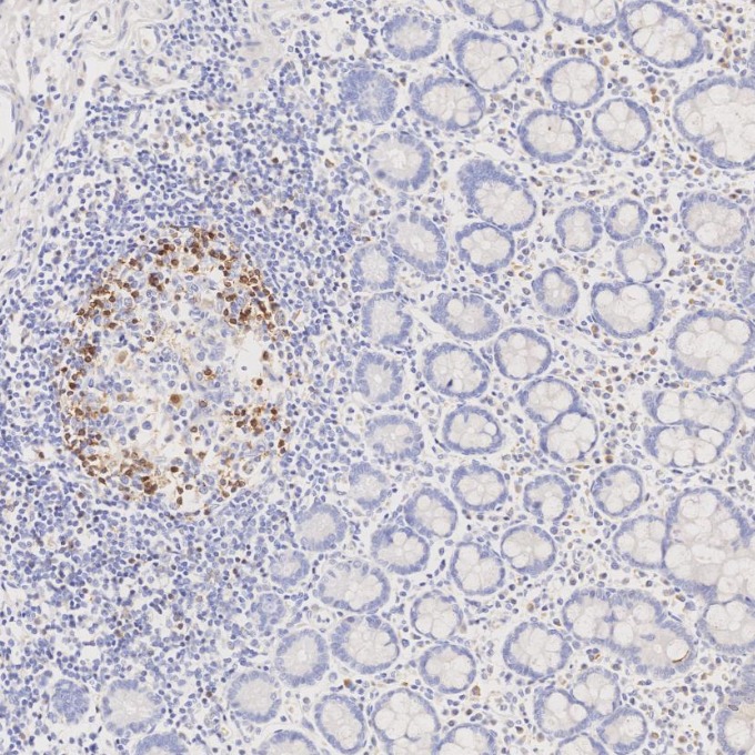
Immunohistochemical analysis of paraffin-embedded human tonsillitis tissue slide using 66220-1-Ig (PD-1/CD279 antibody) at dilution of 1:20000. Heat mediated antigen retrieval with Tris-EDTA buffer (pH 9.0).
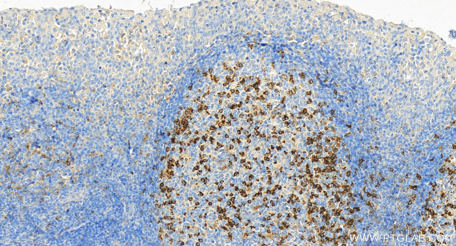
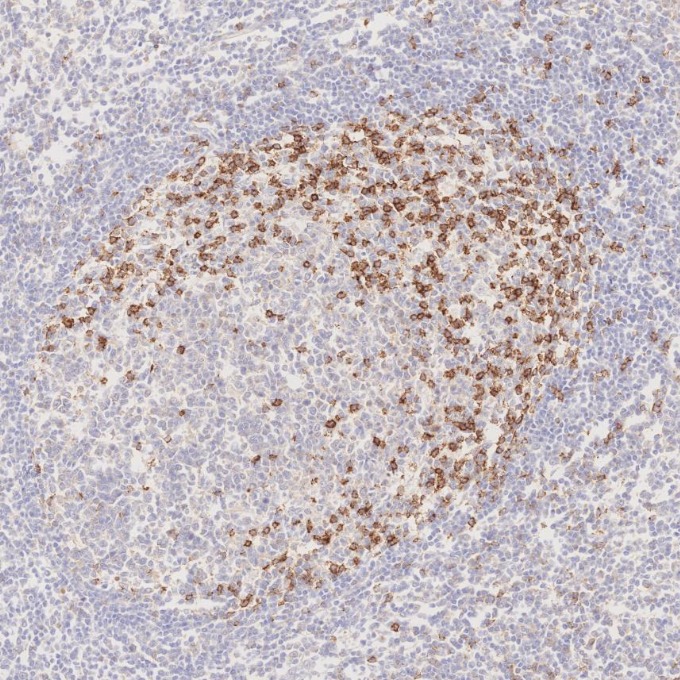
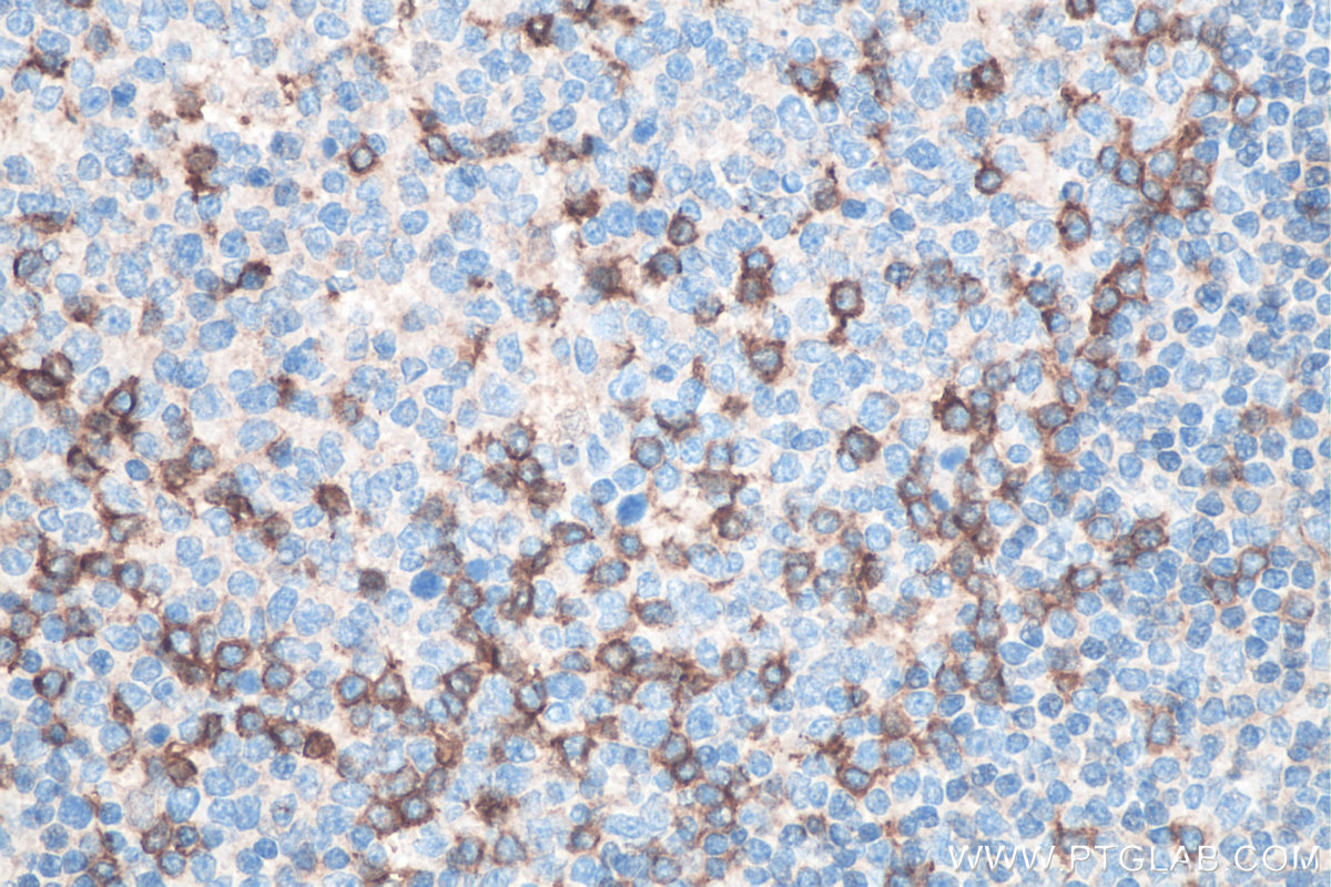
IHC staining of human tonsillitis using 66220-1-Ig
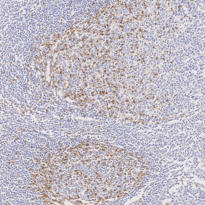
Immunohistochemical analysis of paraffin-embedded human tonsillitis tissue slide using 66220-1-Ig (PD-1/CD279 antibody) at dilution of 1:20000 (under 10x lens). Heat mediated antigen retrieval with Tris-EDTA buffer (pH 9.0).
IHC staining of human tonsillitis using 66220-1-Ig
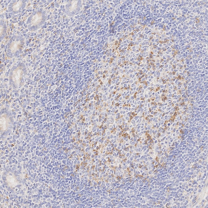
Immunohistochemical analysis of paraffin-embedded human tonsillitis tissue slide using 66220-1-Ig (PD-1/CD279 antibody) at dilution of 1:10000 (under 10x lens). Heat mediated antigen retrieval with Tris-EDTA buffer (pH 9.0).
IHC staining of human tonsillitis using 66220-1-Ig
Immunohistochemical analysis of paraffin-embedded human tonsillitis tissue slide using 66220-1-Ig (PD-1/CD279 antibody) at dilution of 1:10000 (under 40x lens). Heat mediated antigen retrieval with Tris-EDTA buffer (pH 9.0).
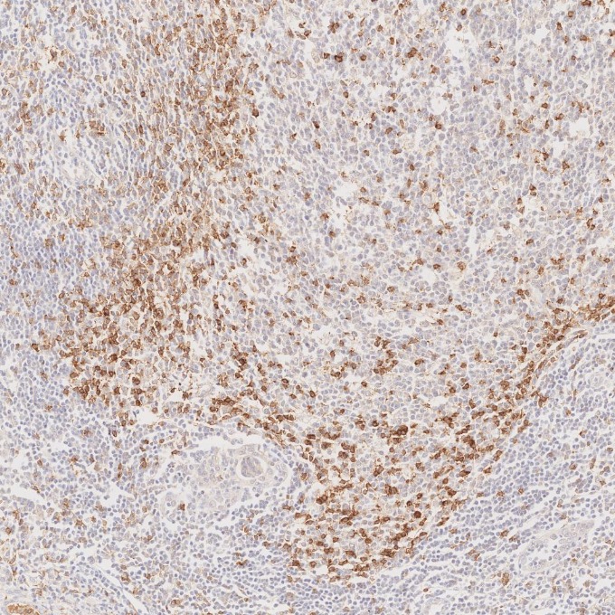
IHC staining of human tonsillitis using 66220-1-Ig
Immunohistochemical analysis of paraffin-embedded human tonsillitis tissue slide using 66220-1-Ig (PD-1/CD279 antibody) at dilution of 1:4000 (under 20x lens). Heat mediated antigen retrieval with Tris-EDTA buffer (pH 9.0).
IHC staining of human tonsillitis using 66220-1-Ig
Immunohistochemical analysis of paraffin-embedded human tonsillitis tissue slide using 66220-1-Ig (PD-1/CD279 antibody) at dilution of 1:4000 (under 20x lens). Heat mediated antigen retrieval with Tris-EDTA buffer (pH 9.0).
IHC-AUTOSTAINER Figures
IHC staining of human appendicitis using 66220-1-Ig
Immunohistochemical analysis of paraffin-embedded human appendicitis tissue slide using 66220-1-Ig (PD-1/CD279 antibody) at dilution of 1:20000. Heat mediated antigen retrieval with Tris-EDTA buffer (pH 9.0). The staining was performed on an autostainer.
IHC staining of human lymphoma using 66220-1-Ig
Immunohistochemical analysis of paraffin-embedded human lymphoma tissue slide using 66220-1-Ig (PD-1/CD279 antibody) at dilution of 1:20000. Heat mediated antigen retrieval with Tris-EDTA buffer (pH 9.0). The staining was performed on an autostainer.
IHC staining of human lymphoma using 66220-1-Ig
Immunohistochemical analysis of paraffin-embedded human lymphoma tissue slide using 66220-1-Ig (PD-1/CD279 antibody) at dilution of 1:20000. Heat mediated antigen retrieval with Tris-EDTA buffer (pH 9.0). The staining was performed on an autostainer.
IHC staining of human tonsillitis using 66220-1-Ig
Immunohistochemical analysis of paraffin-embedded human tonsillitis tissue slide using 66220-1-Ig (PD-1/CD279 antibody) at dilution of 1:20000. Heat mediated antigen retrieval with Tris-EDTA buffer (pH 9.0). The staining was performed on an autostainer.
IF-P Figures
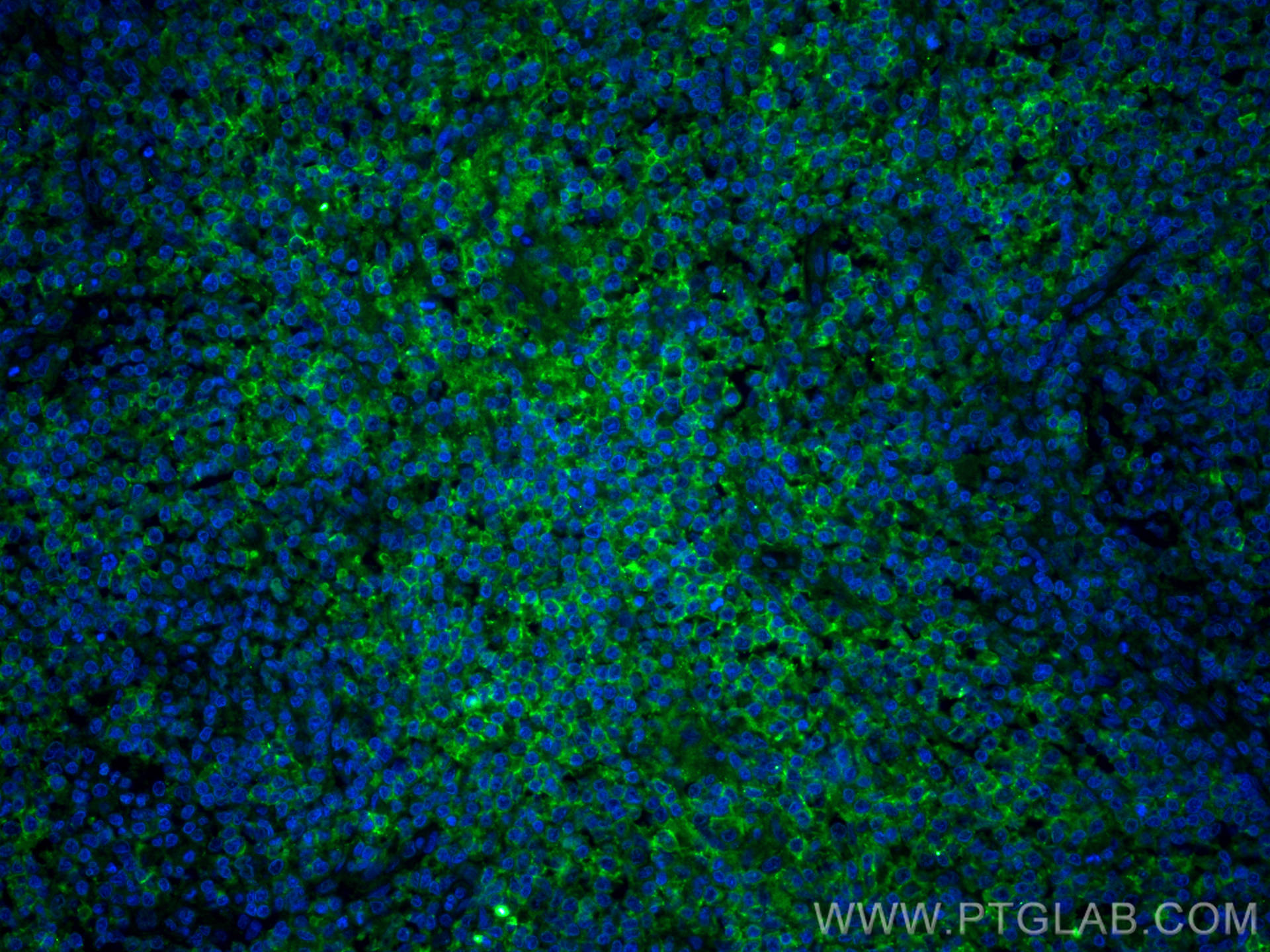
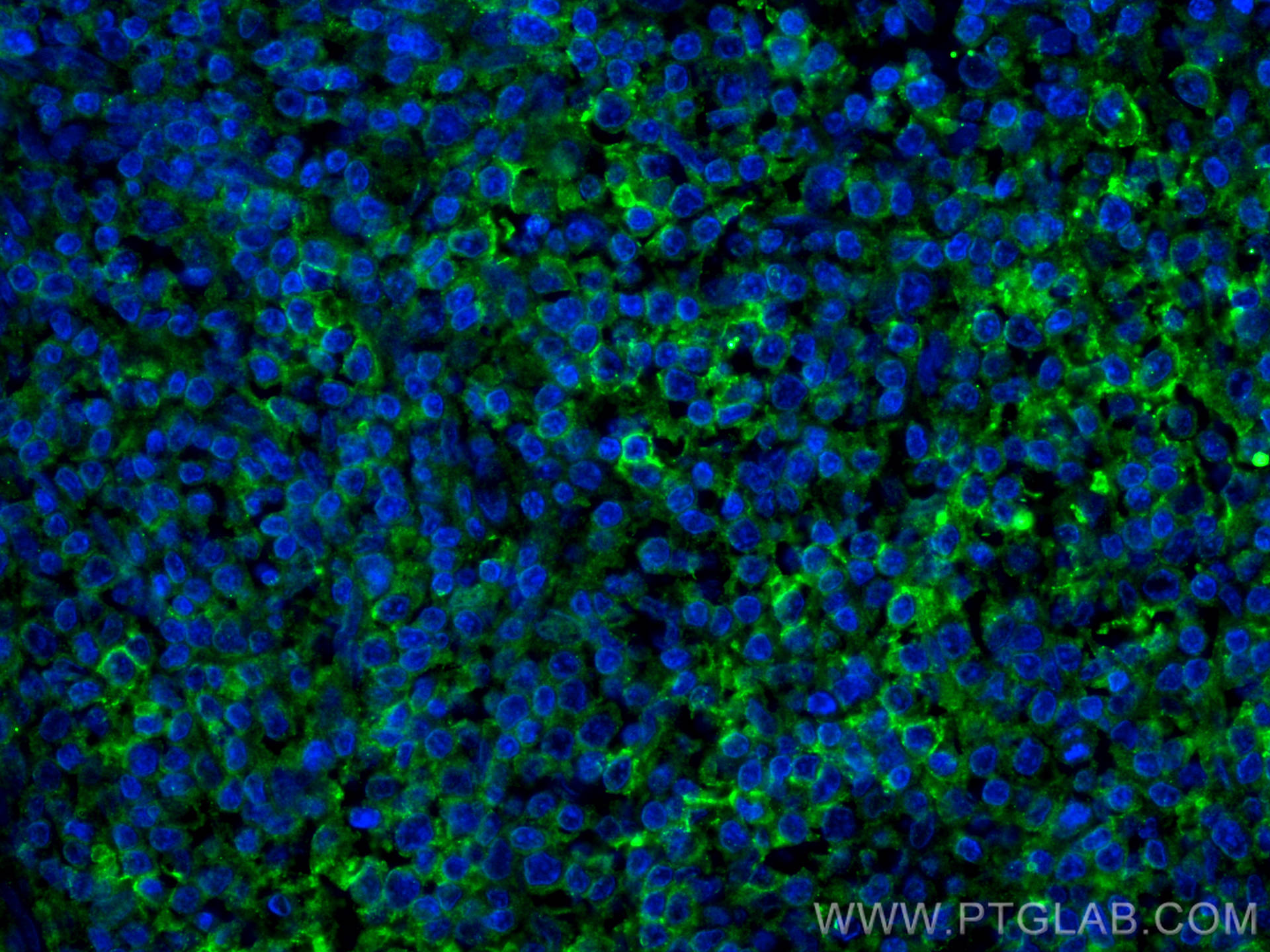
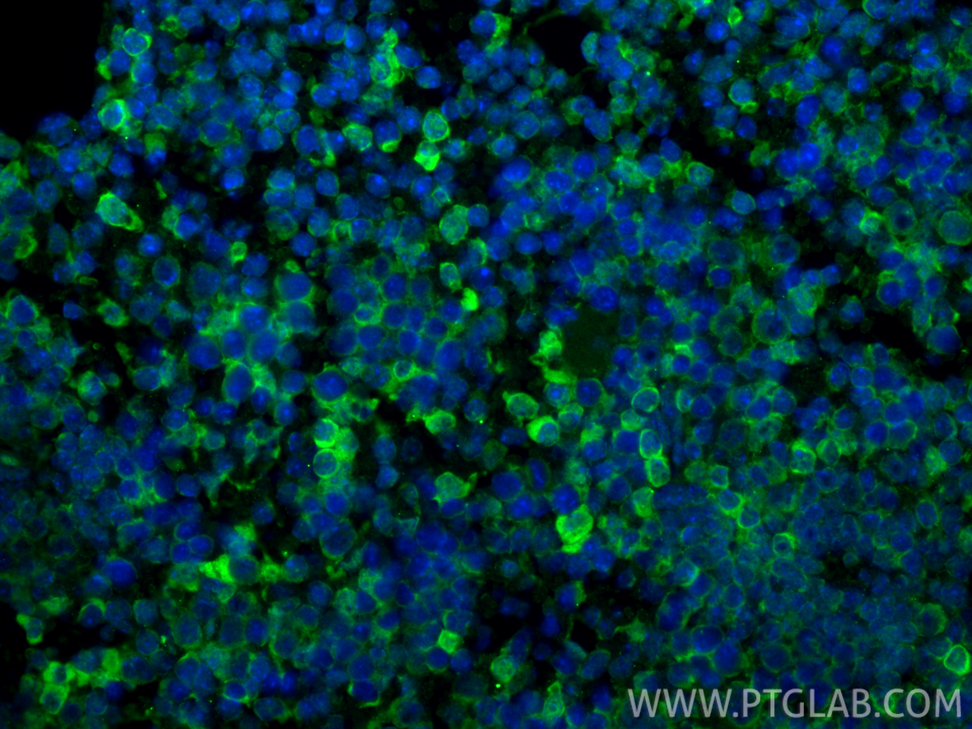
IF Staining of human lymphoma using 66220-1-Ig
Immunofluorescent analysis of (4% PFA) fixed paraffin-embedded human lymphoma tissue using PD-1/CD279 antibody (66220-1-Ig, Clone: 4H4D1 ) at dilution of 1:400 and Multi-rAb CoraLite ® Plus 488-Goat Anti-Mouse Recombinant Secondary Antibody (H+L) (RGAM002). Heat mediated antigen retrieval with Tris-EDTA buffer (pH 9.0).
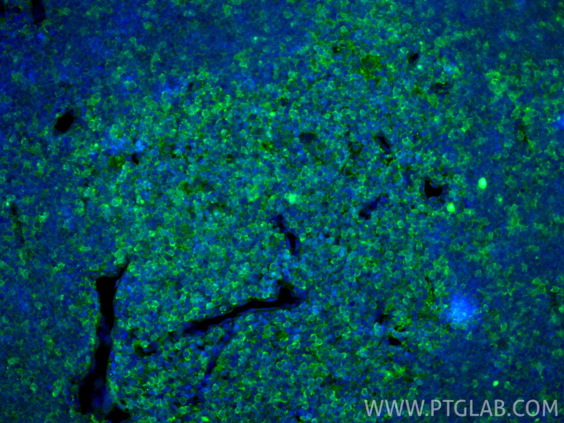
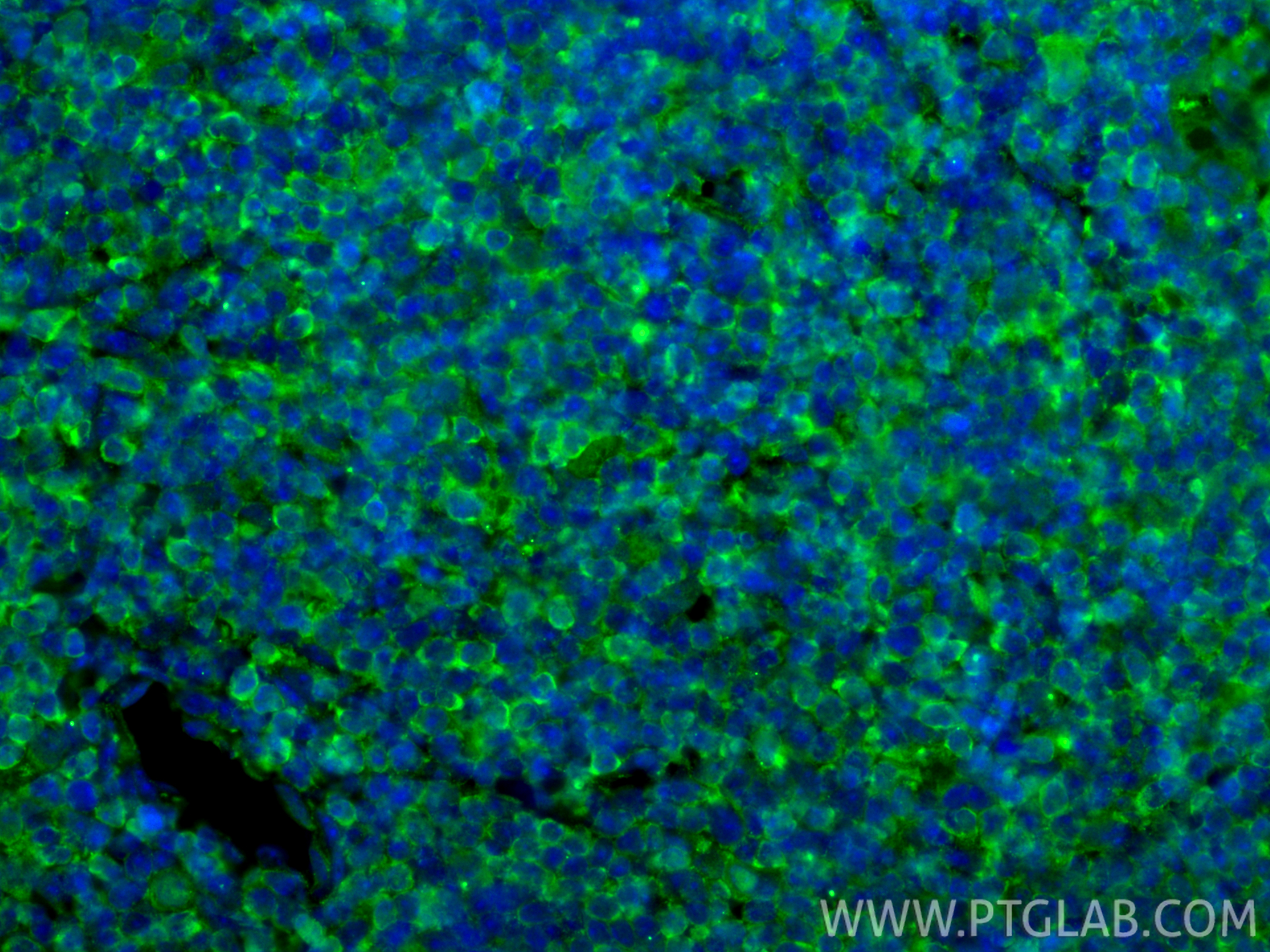
IF Staining of human lymphoma using 66220-1-Ig
Immunofluorescent analysis of (4% PFA) fixed paraffin-embedded human lymphoma tissue using PD-1/CD279 antibody (66220-1-Ig, Clone: 4H4D1 ) at dilution of 1:400 and Multi-rAb CoraLite ® Plus 488-Goat Anti-Mouse Recombinant Secondary Antibody (H+L) (RGAM002). Heat mediated antigen retrieval with Tris-EDTA buffer (pH 9.0).
IF Staining of human tonsillitis using 66220-1-Ig
Immunofluorescent analysis of (4% PFA) fixed paraffin-embedded human tonsillitis tissue using PD-1/CD279 antibody (66220-1-Ig, Clone: 4H4D1 ) at dilution of 1:400 and CoraLite®488-Conjugated Goat Anti-Mouse IgG(H+L) (SA00013-1). Heat mediated antigen retrieval with Tris-EDTA buffer (pH 9.0).
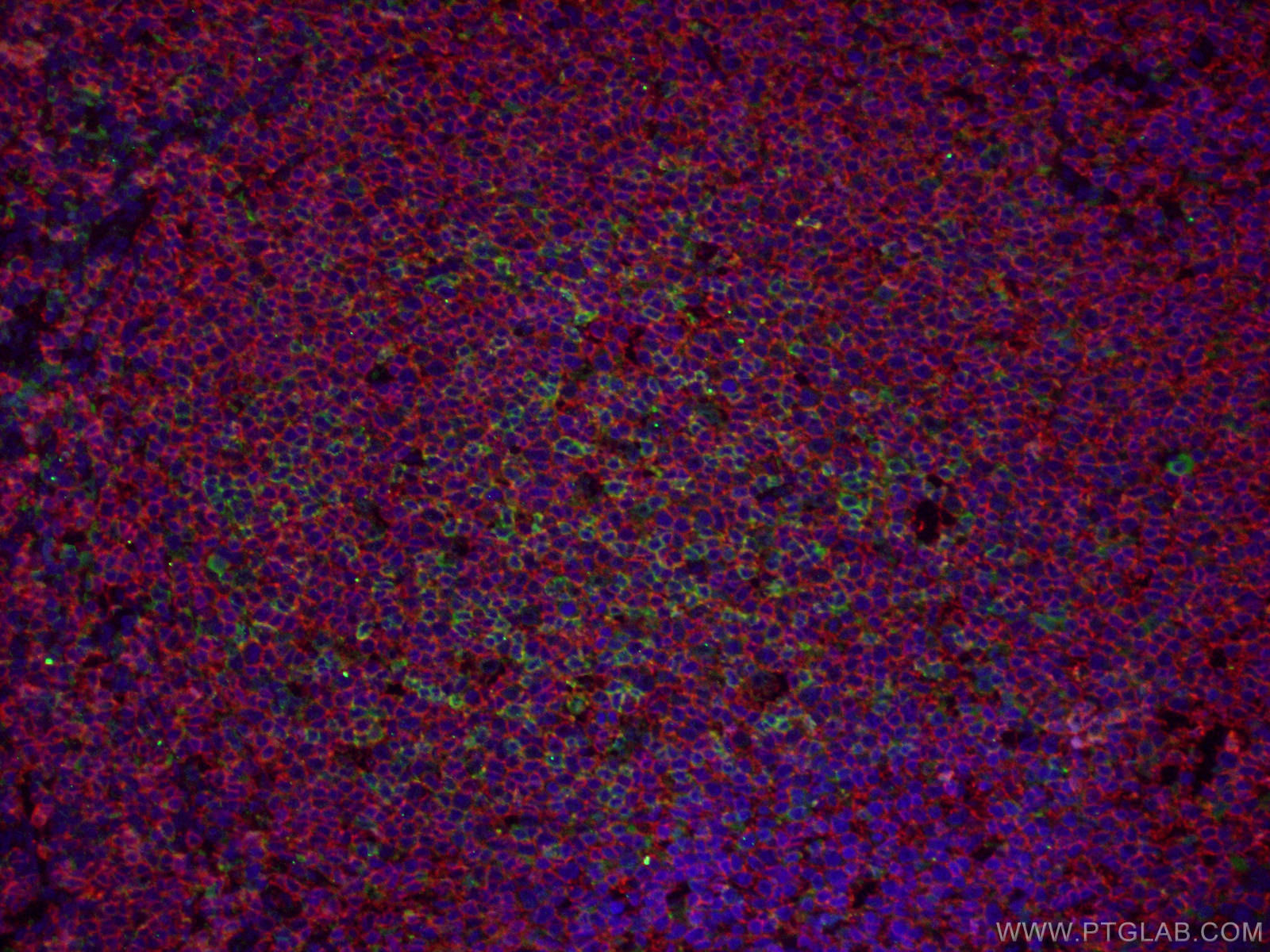
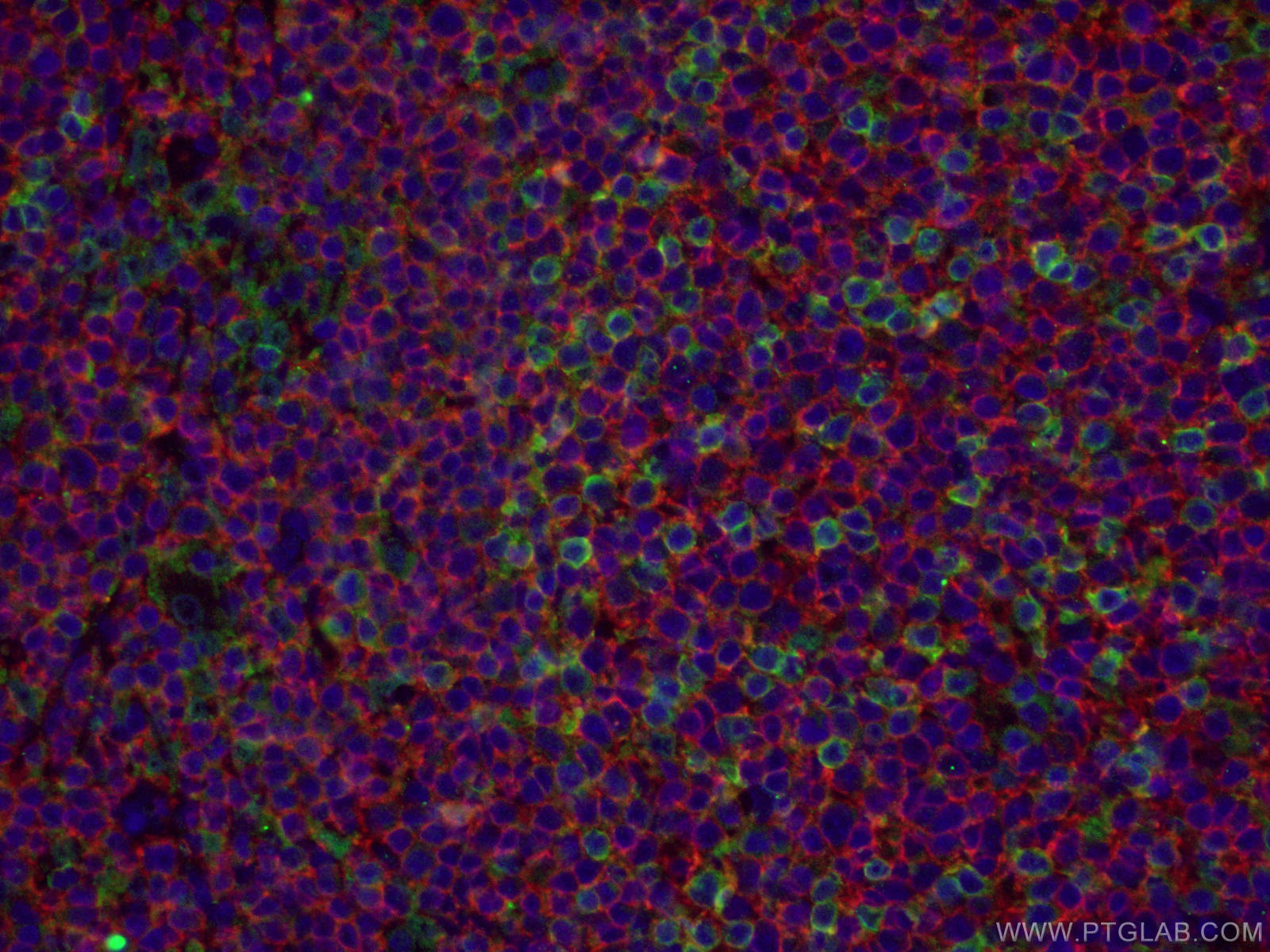
IF Staining of human tonsillitis using 66220-1-Ig
Immunofluorescent analysis of (4% PFA) fixed human tonsillitis tissue using PD-1/CD279 mouse mAb (66220-1-Ig) at dilution of 1:50 and CD20 rabbit pAb (24828-1-AP) at dilution of 1:50, further stained with Alexa Fluor 488-conjugated AffiniPure Goat Anti-Mouse IgG(H+L) for 66220-1-Ig, and Alexa Fluor 594-conjugated AffiniPure Goat Anti-Rabbit IgG (H+L) for 24828-1-AP.
IF Staining of human tonsillitis using 66220-1-Ig
Immunofluorescent analysis of (4% PFA) fixed human tonsillitis tissue using PD-1/CD279 mouse mAb (66220-1-Ig) at dilution of 1:50 and CD20 rabbit pAb (24828-1-AP) at dilution of 1:50, further stained with Alexa Fluor 488-conjugated AffiniPure Goat Anti-Mouse IgG(H+L) for 66220-1-Ig, and Alexa Fluor 594-conjugated AffiniPure Goat Anti-Rabbit IgG (H+L) for 24828-1-AP.
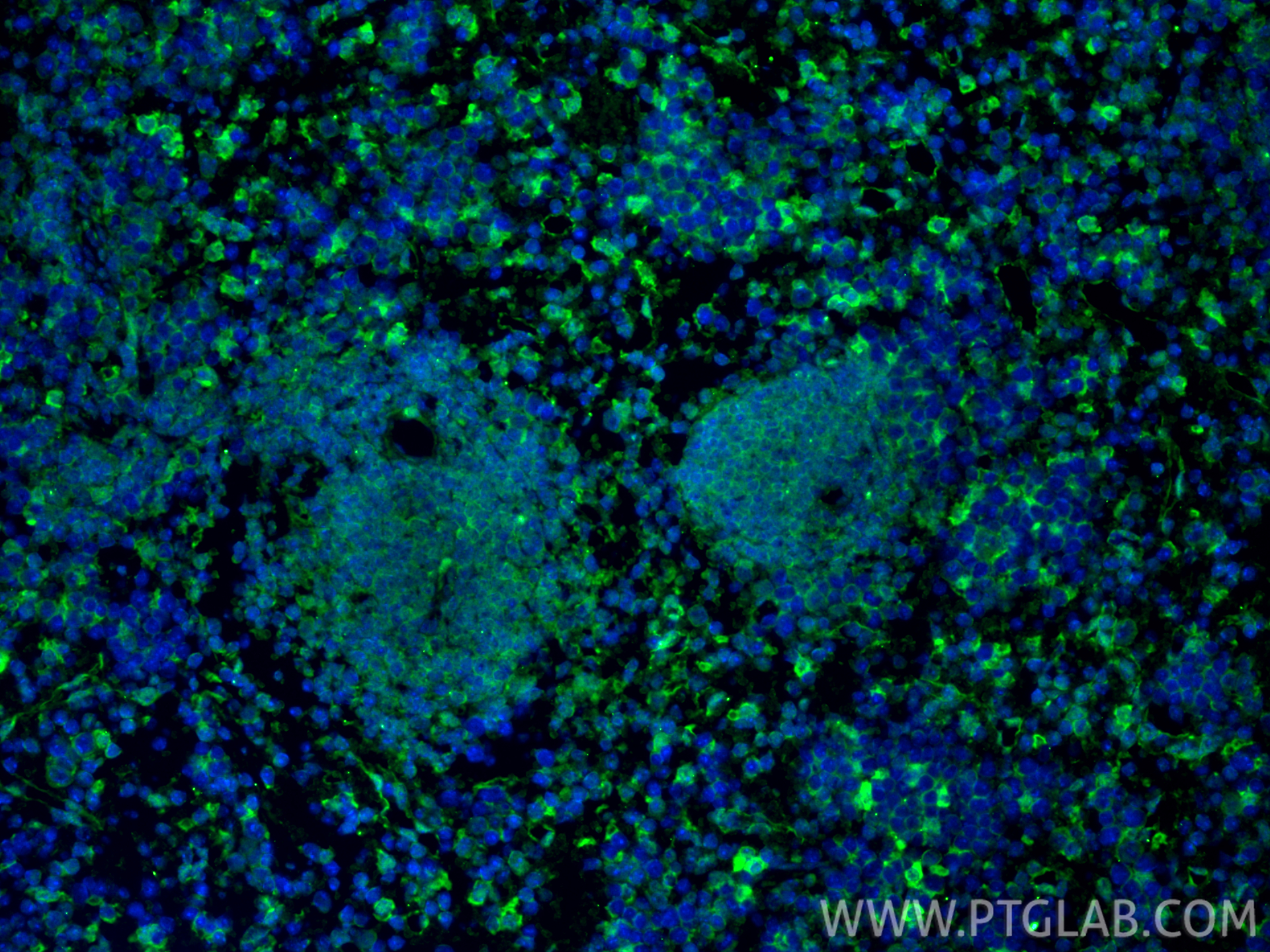
IF Staining of mouse spleen using 66220-1-Ig
Immunofluorescent analysis of (4% PFA) fixed paraffin-embedded mouse spleen tissue using PD-1/CD279 antibody (66220-1-Ig, Clone: 4H4D1 ) at dilution of 1:2000 and CoraLite®488-Conjugated Goat Anti-Mouse IgG(H+L) (SA00013-1). Heat mediated antigen retrieval with Tris-EDTA buffer (pH 9.0).
IF Staining of mouse spleen using 66220-1-Ig
Immunofluorescent analysis of (4% PFA) fixed paraffin-embedded mouse spleen tissue using PD-1/CD279 antibody (66220-1-Ig, Clone: 4H4D1 ) at dilution of 1:2000 and CoraLite®488-Conjugated Goat Anti-Mouse IgG(H+L) (SA00013-1). Heat mediated antigen retrieval with Tris-EDTA buffer (pH 9.0).
IF Staining of mouse thymus using 66220-1-Ig
Immunofluorescent analysis of (4% PFA) fixed paraffin-embedded mouse thymus tissue using PD-1/CD279 antibody (66220-1-Ig, Clone: 4H4D1 ) at dilution of 1:2000 and CoraLite®488-Conjugated Goat Anti-Mouse IgG(H+L) (SA00013-1). Heat mediated antigen retrieval with Tris-EDTA buffer (pH 9.0).
IF Staining of mouse thymus using 66220-1-Ig
Immunofluorescent analysis of (4% PFA) fixed paraffin-embedded mouse thymus tissue using PD-1/CD279 antibody (66220-1-Ig, Clone: 4H4D1 ) at dilution of 1:2000 and CoraLite®488-Conjugated Goat Anti-Mouse IgG(H+L) (SA00013-1). Heat mediated antigen retrieval with Tris-EDTA buffer (pH 9.0).