验证数据展示

经过测试的应用
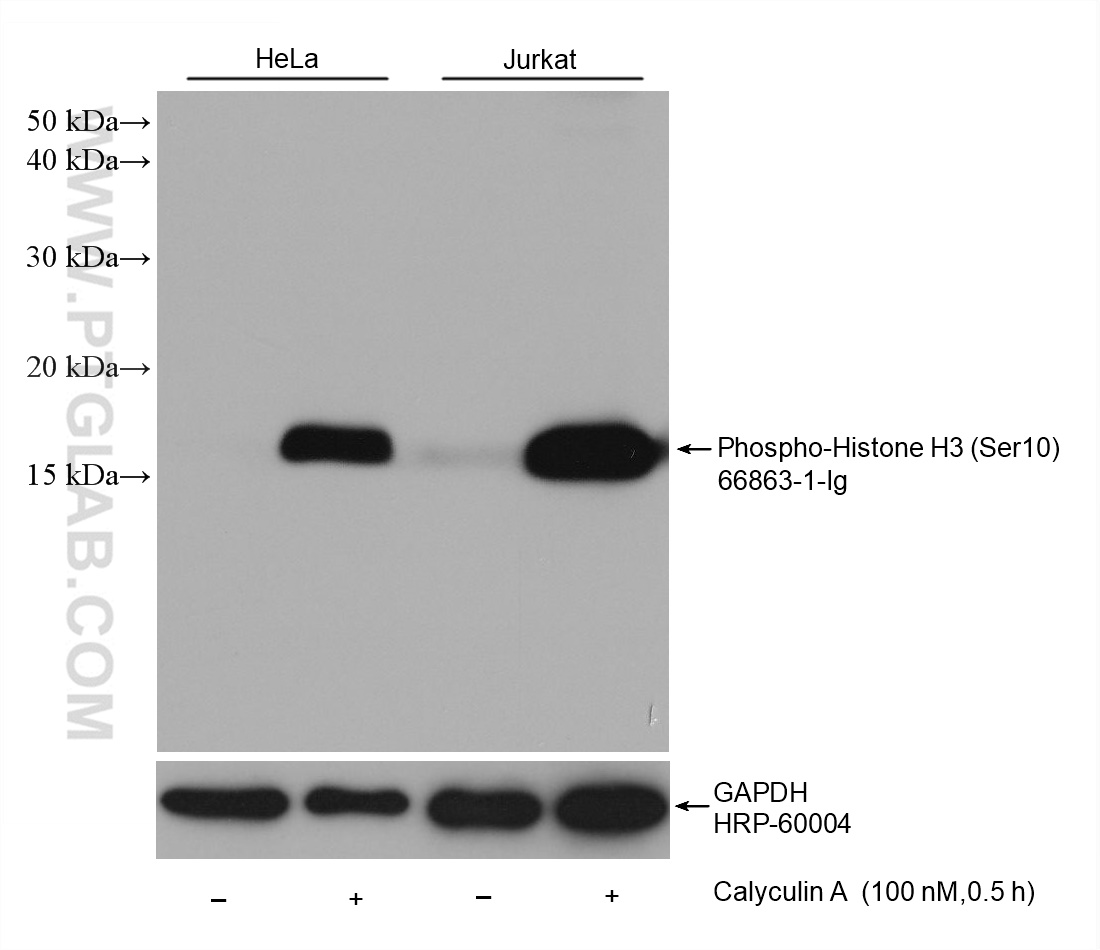
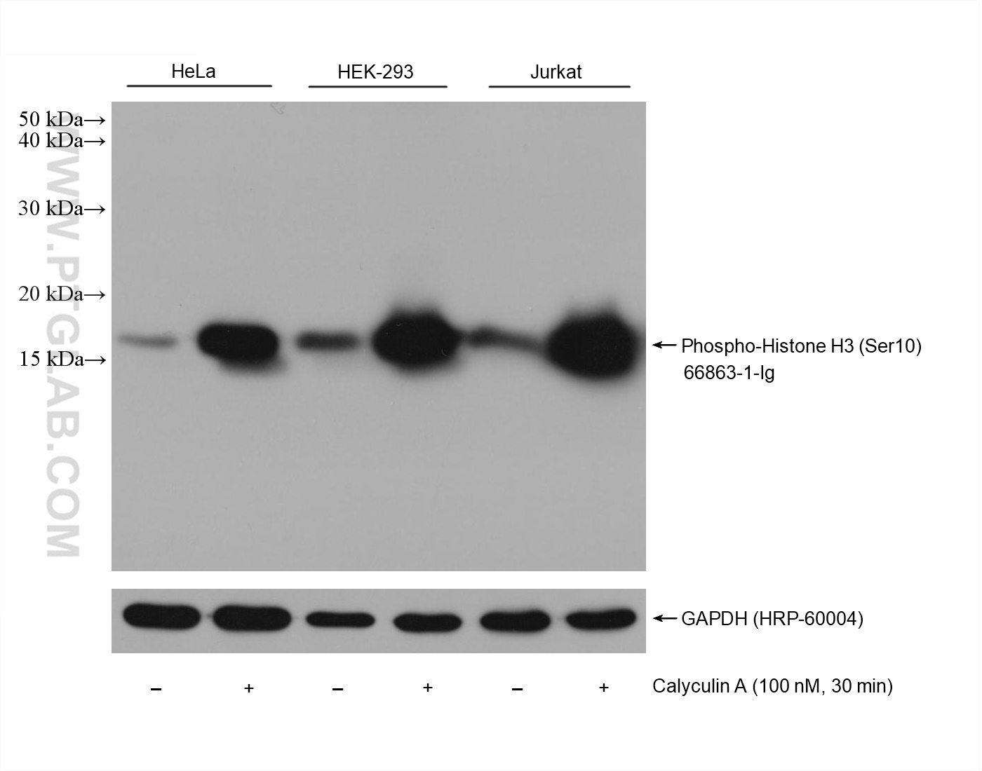
| Positive WB detected in | HeLa cells, HEK-293 cells, Jurkat cells |
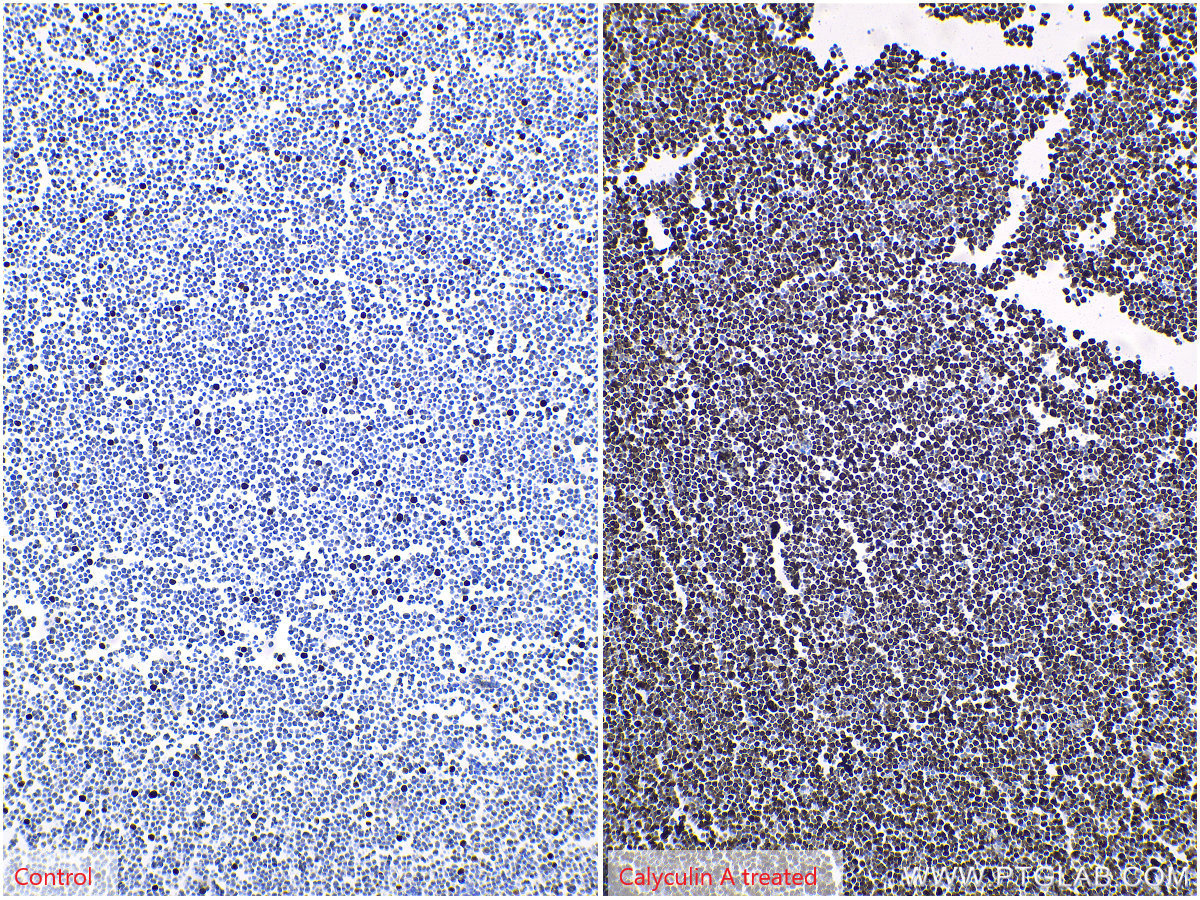
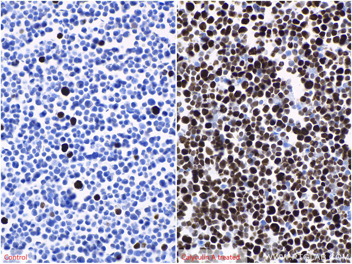
| Positive IHC detected in | Jurkat cells Note: suggested antigen retrieval with TE buffer pH 9.0; (*) Alternatively, antigen retrieval may be performed with citrate buffer pH 6.0 |
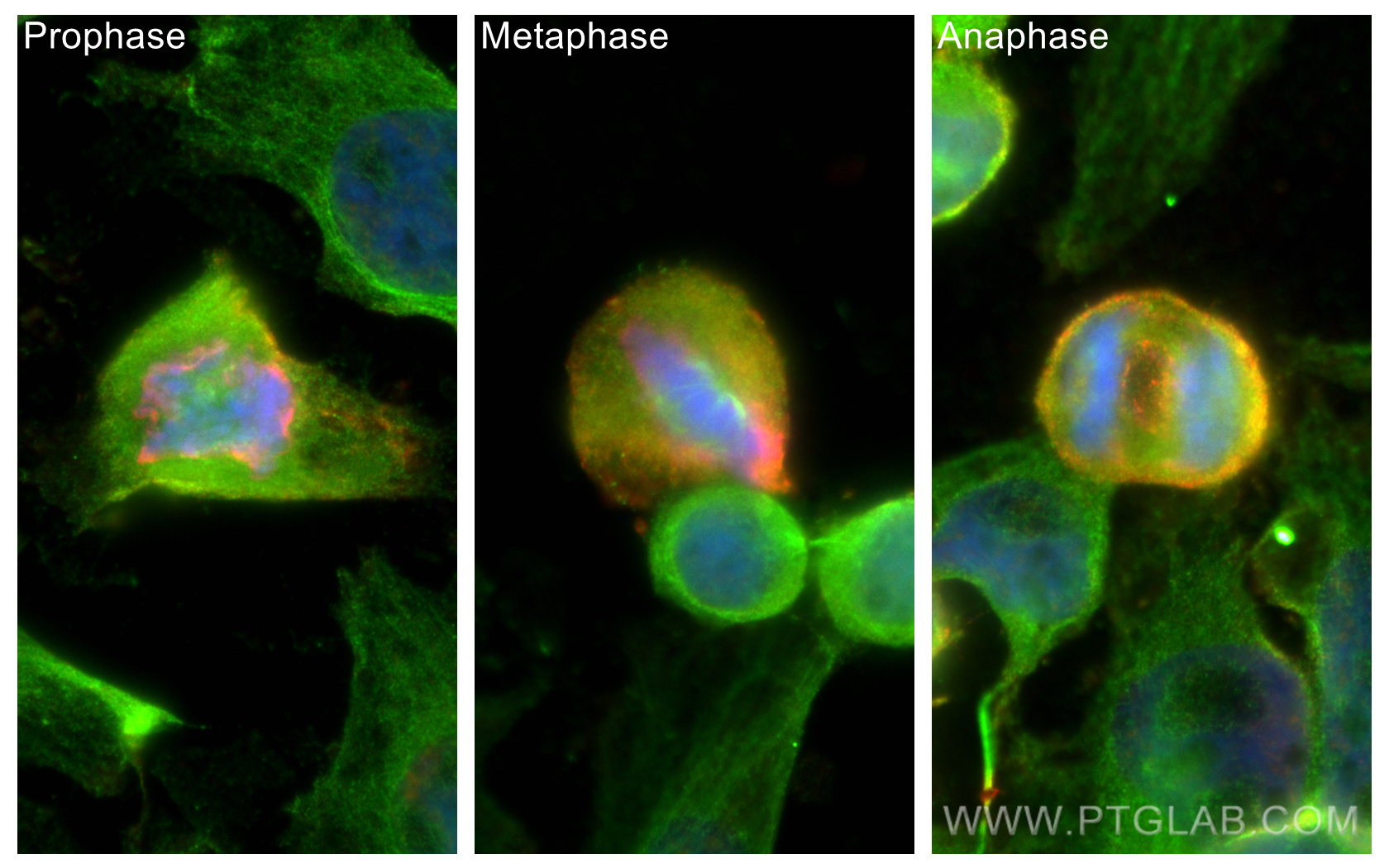
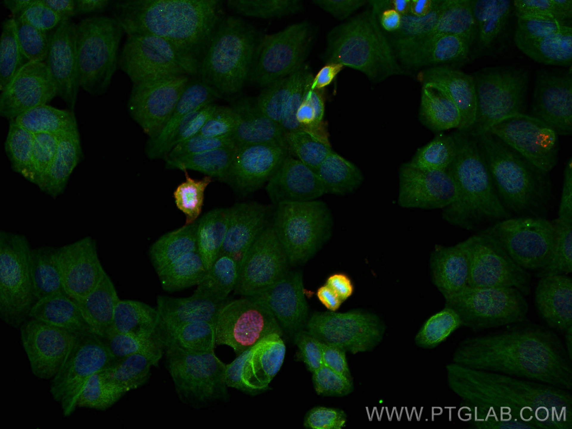
| Positive IF/ICC detected in | HeLa cells, MCF-7 cells |
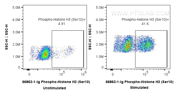
| Positive FC (Intra) detected in | nocodazole treated HeLa cells |
推荐稀释比
| 应用 | 推荐稀释比 |
|---|---|
| Western Blot (WB) | WB : 1:5000-1:50000 |
| Immunohistochemistry (IHC) | IHC : 1:1000-1:4000 |
| Immunofluorescence (IF)/ICC | IF/ICC : 1:500-1:2000 |
| Flow Cytometry (FC) (INTRA) | FC (INTRA) : 0.25 ug per 10^6 cells in a 100 µl suspension |
| It is recommended that this reagent should be titrated in each testing system to obtain optimal results. | |
| Sample-dependent, Check data in validation data gallery. | |
产品信息
66863-1-Ig targets Phospho-Histone H3 (Ser10) in WB, IHC, IF/ICC, FC (Intra), ELISA applications and shows reactivity with human samples.
| 经测试应用 | WB, IHC, IF/ICC, FC (Intra), ELISA Application Description |
| 文献引用应用 | WB, IHC, IF |
| 经测试反应性 | human |
| 文献引用反应性 | human, mouse, rat, chicken |
| 免疫原 |
Peptide 种属同源性预测 |
| 宿主/亚型 | Mouse / IgG1 |
| 抗体类别 | Monoclonal |
| 产品类型 | Antibody |
| 全称 | histone cluster 1, H3a |
| 别名 | H3, H3 P-S10, H3S10, H3S10Ph, Histone H3 (Ser10) |
| 计算分子量 | 15 kDa |
| 观测分子量 | 15-17 kDa |
| GenBank蛋白编号 | NM_003529 |
| 基因名称 | HIST1H3A |
| Gene ID (NCBI) | 8350 |
| RRID | AB_2882201 |
| 偶联类型 | Unconjugated |
| 形式 | Liquid |
| 纯化方式 | Protein G purification |
| UNIPROT ID | P68431 |
| 储存缓冲液 | PBS with 0.02% sodium azide and 50% glycerol, pH 7.3. |
| 储存条件 | Store at -20°C. Stable for one year after shipment. Aliquoting is unnecessary for -20oC storage. |
背景介绍
Phospho-histone-H3 (PHH3) is a core histone protein, which in its phosphorylated state forms the principal constituents of eukaryotic chromatin, with histone H3 being phosphorylated at serine (Ser) 10 or Ser28 as well as its phosphorylation of Ser10 being strongly correlated with the late G2 to M-phase transition in mammalian mitotic cells. On the basis of previous research, a few cell line- and animal model-based researches have displayed an increase in phosphorylation of histone H3 at Ser10 (H3S10ph), the only histone marker that is involved in carcinogenesis and cellular transformation. Histone H3 phosphorylation on serine-10 is specific to mitosis and phosphorylated histone H3 (PHH3) proliferation markers (as counts defined per area or as indices defined per cell numbers) are increasingly being used to evaluate proliferation in various tumors.
实验方案
| Product Specific Protocols | |
|---|---|
| IF protocol for Phospho-Histone H3 (Ser10) antibody 66863-1-Ig | Download protocol |
| IHC protocol for Phospho-Histone H3 (Ser10) antibody 66863-1-Ig | Download protocol |
| WB protocol for Phospho-Histone H3 (Ser10) antibody 66863-1-Ig | Download protocol |
| Standard Protocols | |
|---|---|
| Click here to view our Standard Protocols |
发表文章
| Species | Application | Title |
|---|---|---|
Mol Cancer Deubiquitination of CDC6 by OTUD6A promotes tumour progression and chemoresistance | ||
Natl Sci Rev Epigenomic landscapes during prefrontal cortex development and aging in rhesus | ||
Nat Commun Medium from human iPSC-derived primitive macrophages promotes adult cardiomyocyte proliferation and cardiac regeneration | ||
J Nanobiotechnology Hepatocyte-derived tissue extracellular vesicles safeguard liver regeneration and support regenerative therapy | ||
Mater Today Bio Aptamer-functionalized exosomes combined with doxorubicin suppress GBM progression and enhance chemoradiosensitivity by promoting pyroptosis. |