Recombinant Mouse MSR1 protein (rFc Tag)
种属
Mouse
纯度
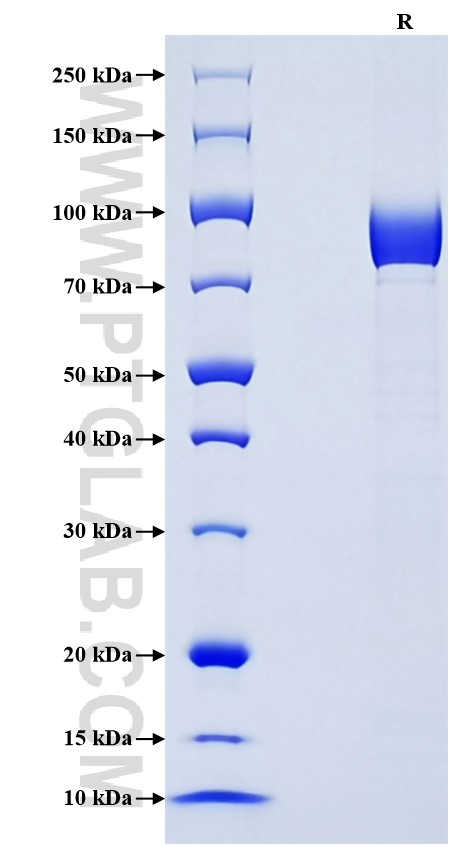
>90 %, SDS-PAGE
标签
N-rFc
生物活性
未测试
验证数据展示
产品信息
| 纯度 | >90 %, SDS-PAGE |
| 内毒素 | <0.1 EU/μg protein, LAL method |
| 生物活性 |
Not tested |
| 来源 | HEK293-derived Mouse MSR1 protein Gln79-Ser458 (Accession# P30204-1) with a rabbit IgG Fc tag at the N-terminus. |
| 基因ID | 20288 |
| 蛋白编号 | P30204-1 |
| 预测分子量 | 68.8 kDa |
| SDS-PAGE | 75-100 kDa, reducing (R) condition |
| 组分 | Lyophilized from 0.22 μm filtered solution in PBS, pH 7.4. Normally 5% trehalose and 5% mannitol are added as protectants before lyophilization. |
| 复溶 | Briefly centrifuge the tube before opening. Reconstitute at 0.1-0.5 mg/mL in sterile water. |
| 储存条件 |
It is recommended that the protein be aliquoted for optimal storage. Avoid repeated freeze-thaw cycles.
|
| 运输条件 | The product is shipped at ambient temperature. Upon receipt, store it immediately at the recommended temperature. |
背景信息
Macrophage scavenger receptor 1 (MSR1), also known as Scara1, is a type II transmembrane glycoprotein primarily expressed by microglia in the central nervous system (CNS) of mice. MSR1 plays a significant role in host defense against bacterial infections and has been implicated in the control of viral infections through autophagy. As an important phagocytic receptor, MSR1 can mediate the uptake of a wide range of ligands, including low-density lipoproteins, bacterial pathogens, and dead cells. This function is crucial for the clearance of misfolded protein aggregates, which may include prions, similar to its role in AD with Aβ. MSR1 contributes to inflammatory disorders and cancer. It has been shown to facilitate cellular entry of vesicular stomatitis virus (VSV) in an LDLR family-dependent manner and interacts with VSV glycoprotein G via its extracellular domains. Additionally, MSR1-mediated uptake of saturated fatty acids can activate JNK signaling and induce Tnf-α and Il-6 expression, leading to a phenotype switch from anti-inflammatory to pro-inflammatory.
参考文献:
1. Jack Gudgeon. et al. (2022). Front Immunol. 13:1012002.
2. Long Yang. et al. (2020). Commun Biol. 3(1):556.
3. Bei Li. et al. (2021). J Mol Med (Berl). 99(6):877-887.
4. Duomeng Yang. et al. (2021). iScience. 24(6):102678.