验证数据展示
")
")
")
")
")
")
")
")
")
")
")
产品信息
22787-1-PBS targets MCT4 in WB, IHC, IF/ICC, FC (Intra), ELISA applications and shows reactivity with human, mouse, rat samples.
| 经测试应用 | WB, IHC, IF/ICC, FC (Intra), ELISA Application Description |
| 经测试反应性 | human, mouse, rat |
| 免疫原 |
CatNo: Ag18788 Product name: Recombinant human SLC16A3 protein Source: e coli.-derived, PGEX-4T Tag: GST Domain: 402-465 aa of BC112269 Sequence: LLLGNFFCIRKKPKEPQPEVAAAEEEKLHKPPADSGVDLREVEHFLKAEPEKNGEVVHTPETSV 种属同源性预测 |
| 宿主/亚型 | Rabbit / IgG |
| 抗体类别 | Polyclonal |
| 产品类型 | Antibody |
| 全称 | solute carrier family 16, member 3 (monocarboxylic acid transporter 4) |
| 别名 | MCT-4, SLC16A3, MCT 4, MCT3 |
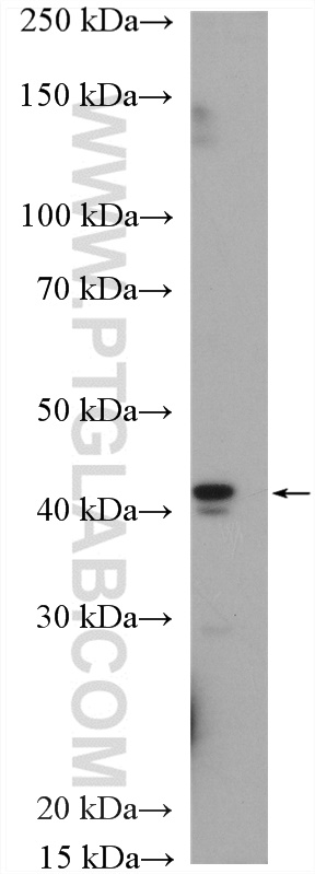
| 计算分子量 | 465 aa, 49 kDa |
| 观测分子量 | 38-42 kDa |
| GenBank蛋白编号 | BC112269 |
| 基因名称 | MCT4 |
| Gene ID (NCBI) | 9123 |
| RRID | AB_11182479 |
| 偶联类型 | Unconjugated |
| 形式 | Liquid |
| 纯化方式 | Antigen affinity purification |
| UNIPROT ID | O15427 |
| 储存缓冲液 | PBS only, pH 7.3. |
| 储存条件 | Store at -80°C. The product is shipped with ice packs. Upon receipt, store it immediately at -80°C |
背景介绍
The monocarboxylate transporter 4 (MCT4, also known as SLC16A3) is involved in the transportation of metabolically important monocarboxylates such as lactate, pyruvate, acetate and ketone bodies. It is widely expressed, particularly strongly in glycolytic tissues such as white skeletal muscle fibres, astrocytes, white blood cells, chondrocytes and some mammalian cell lines. MCT4 is also linked to tumor biology because it mediates lactate transport across membranes resulting in antiapoptotic effects.