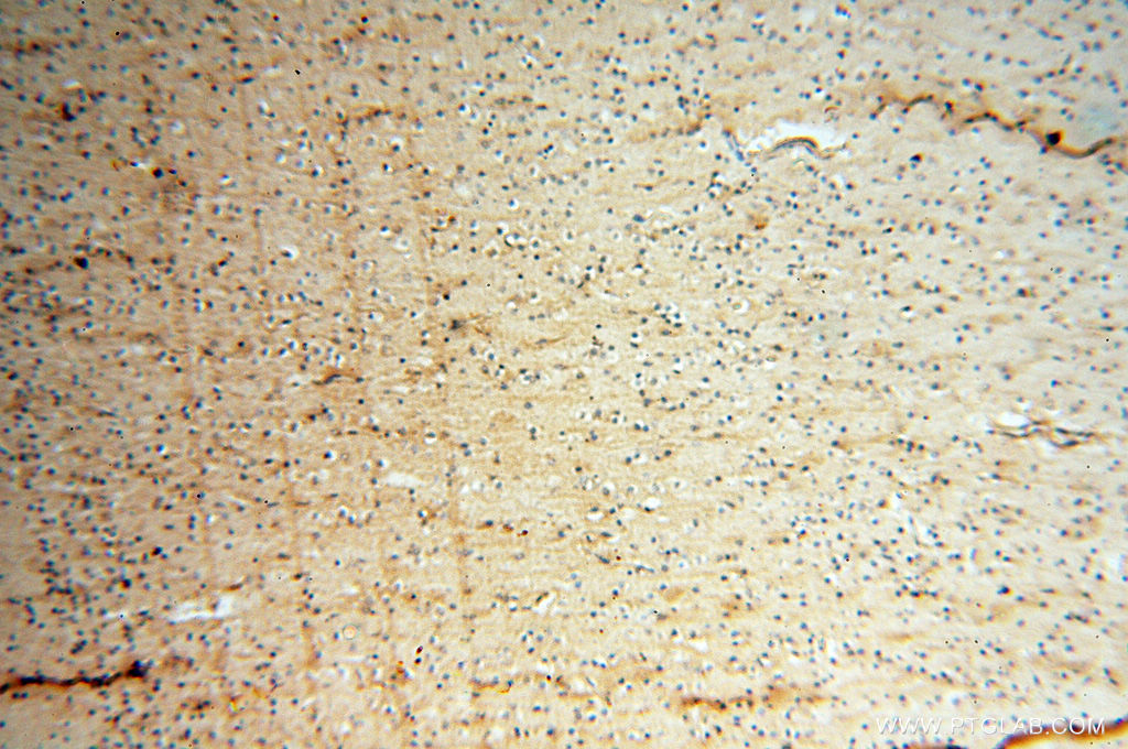
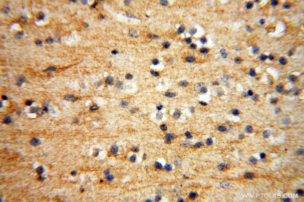
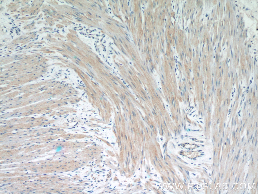
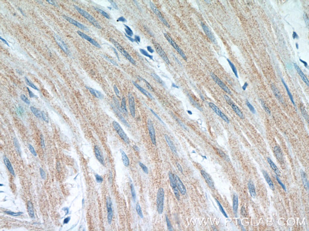
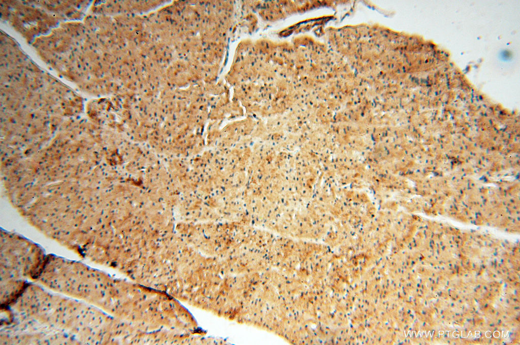
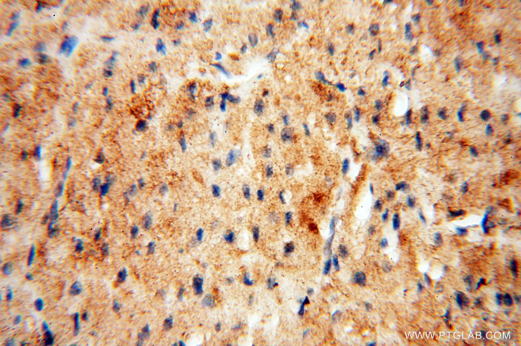
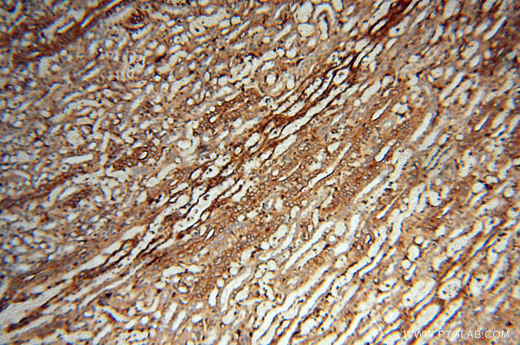
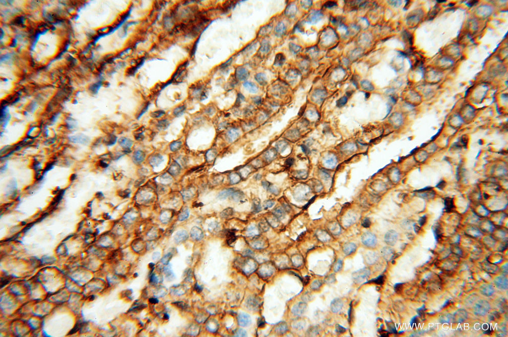
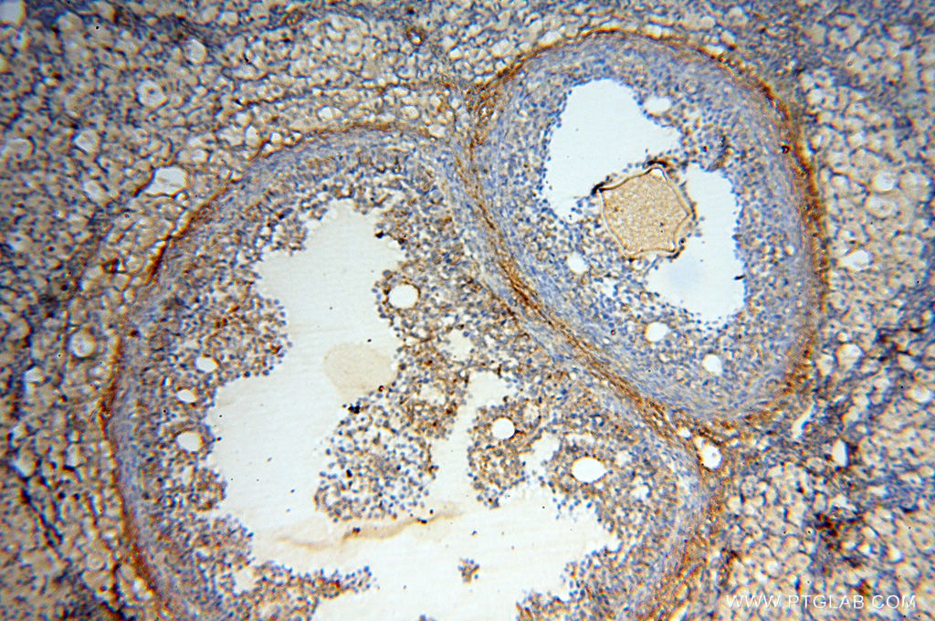
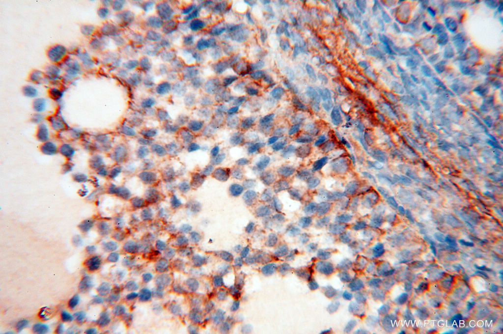
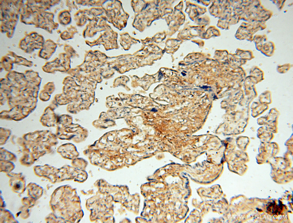
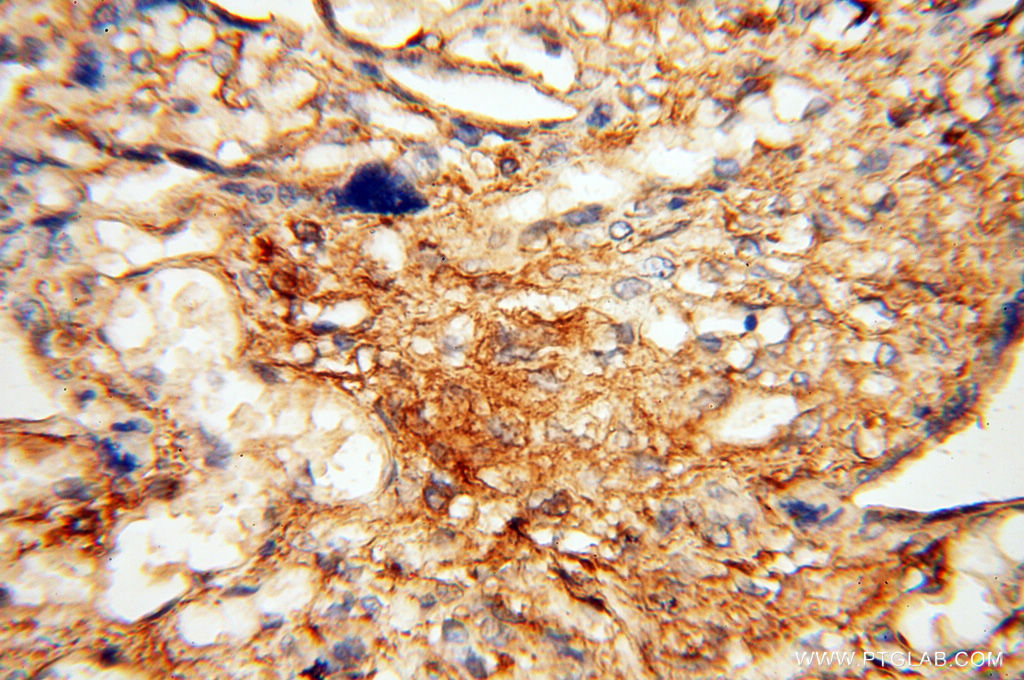
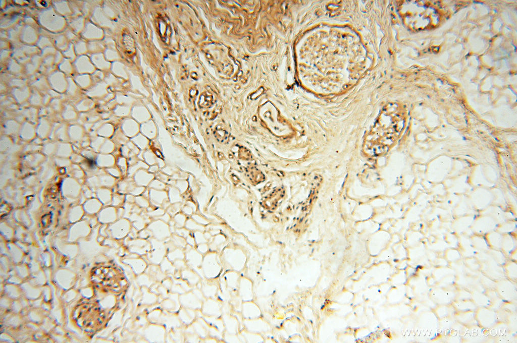
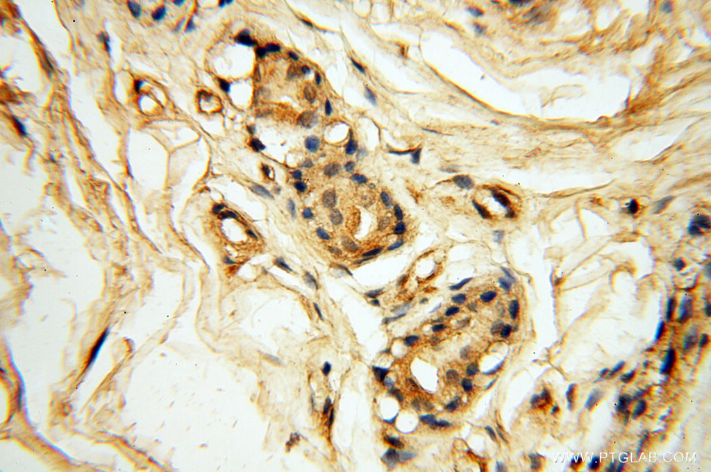
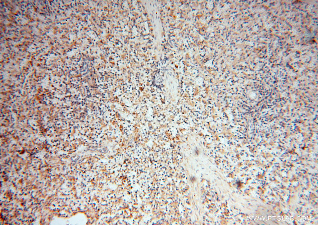
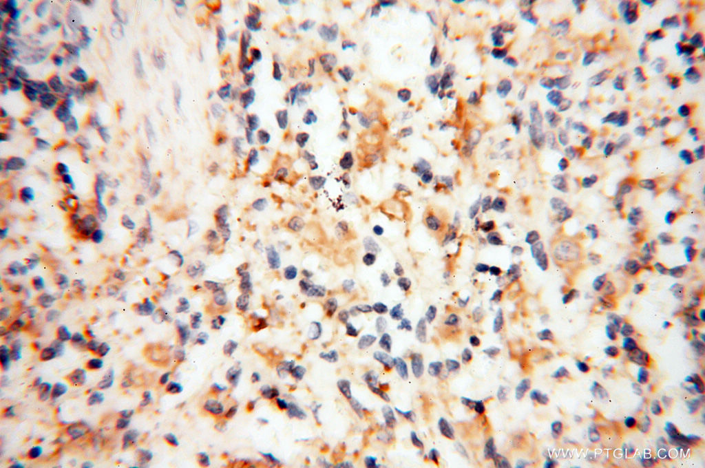
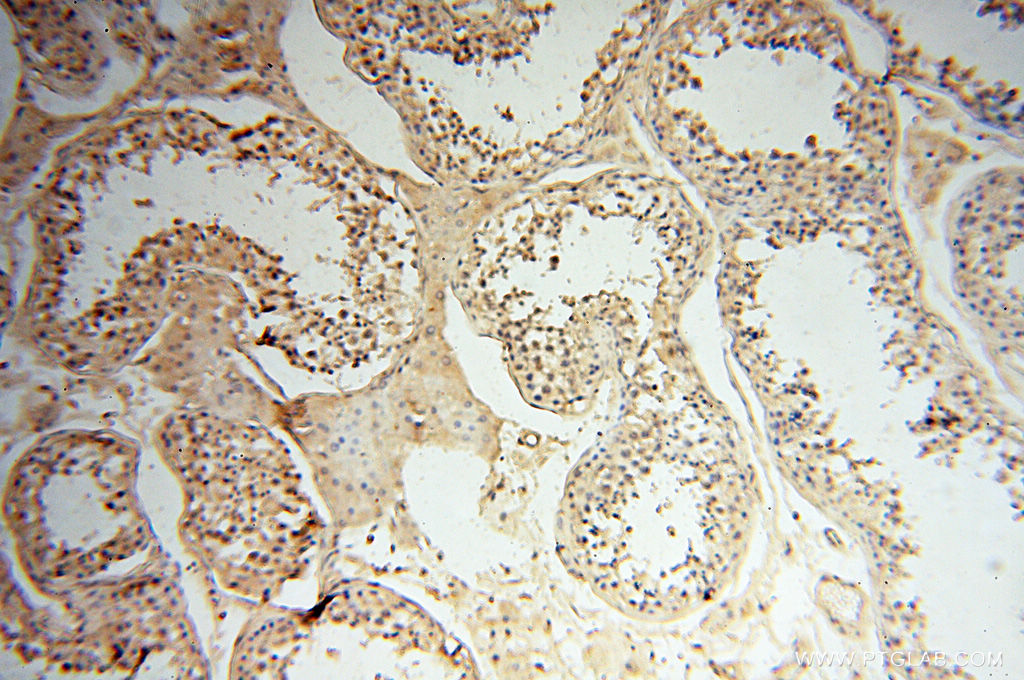
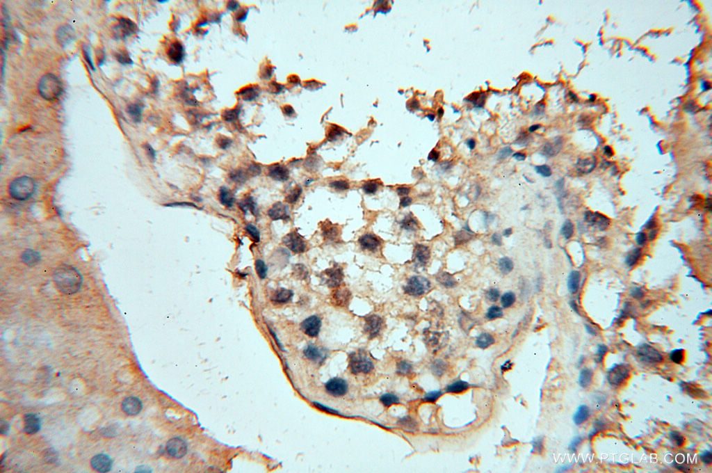
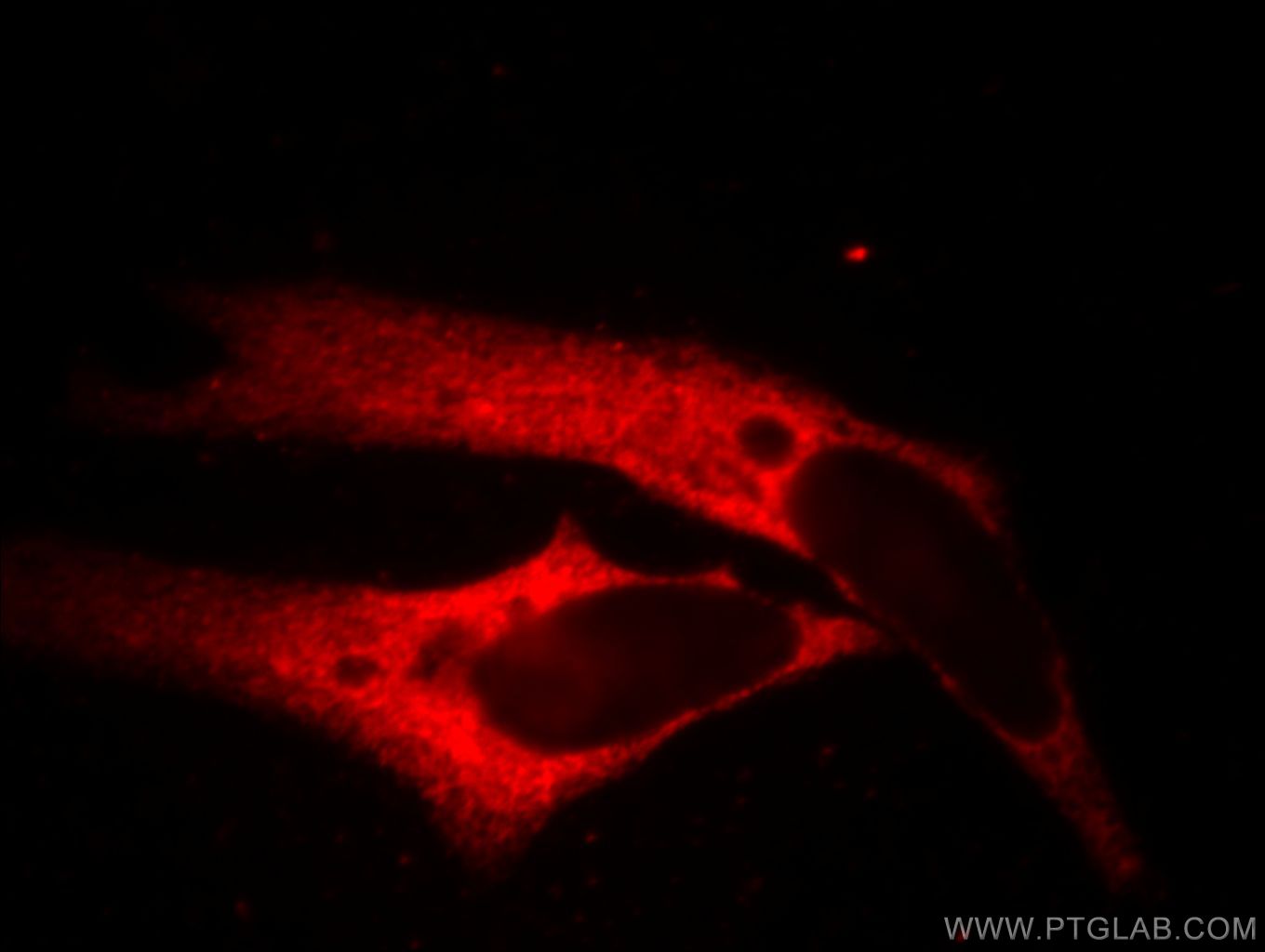
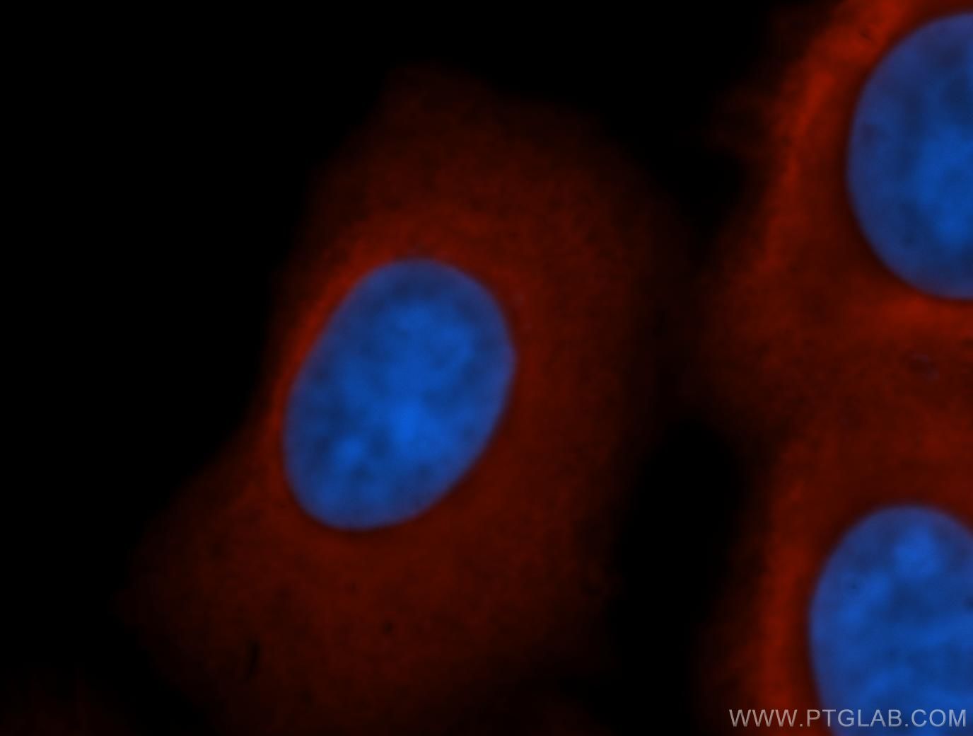
验证数据展示
发表文章中的应用
| WB | See 7 publications below |
产品信息
60008-2-Ig targets beta actin in WB, ELISA applications and shows reactivity with human samples.
| 经测试应用 | ELISA Application Description |
| 文献引用应用 | WB |
| 经测试反应性 | human |
| 文献引用反应性 | human, mouse |
| 免疫原 |
CatNo: Ag0297 Product name: Recombinant human beta actin protein Source: e coli.-derived, PGEX-4T Tag: GST Domain: 14-167 aa of BC002409 Sequence: SGMCKAGFAGDDAPRAVFPSIVGRPRHQGVMVGMGQKDSYVGDEAQSKRGILTLKYPIEHGIVTNWDDMEKIWHHTFYNELRVAPEEHPVLLTEAPLNPKANREKMTQIMFETFNTPAMYVAIQAVLSLYASGRTTGIVMDSGDGVTHTVPIYE 种属同源性预测 |
| 宿主/亚型 | Mouse / IgM |
| 抗体类别 | Monoclonal |
| 产品类型 | Antibody |
| 全称 | actin, beta |
| 别名 | |
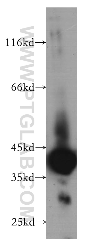
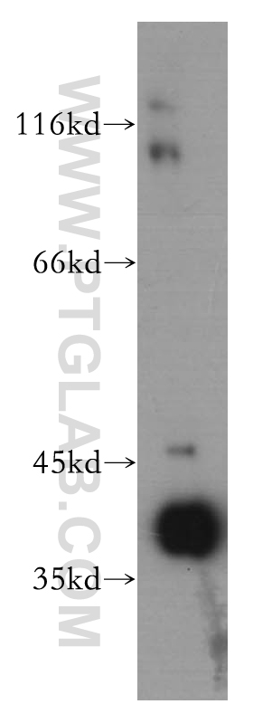
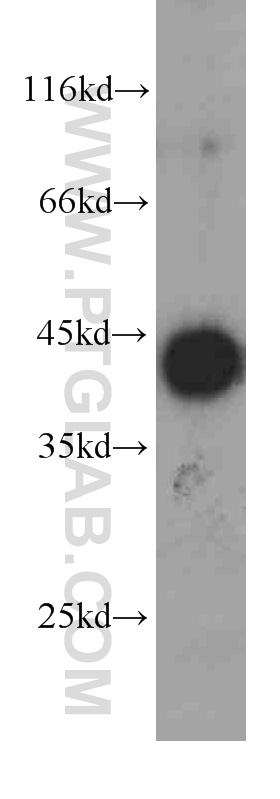
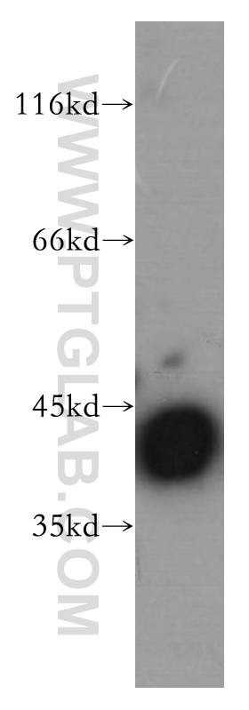
| 计算分子量 | 375 aa, 42 kDa |
| 观测分子量 | 45 kDa |
| GenBank蛋白编号 | BC002409 |
| 基因名称 | Beta Actin |
| Gene ID (NCBI) | 60 |
| 偶联类型 | Unconjugated |
| 形式 | Liquid |
| 纯化方式 | Caprylic Acid/Ammonium Sulfate Precipitation |
| UNIPROT ID | P60709 |
| 储存缓冲液 | PBS with 0.02% sodium azide and 50% glycerol, pH 7.3. |
| 储存条件 | Store at -20°C. Stable for one year after shipment. Aliquoting is unnecessary for -20oC storage. |
背景介绍
Beta actin, also named as ACTB and F-Actin, belongs to the actin family. Actins are highly conserved globular proteins that are involved in various types of cell motility and are ubiquitously expressed in all eukaryotic cells. At least six isoforms of actins are known in mammals and other vertebrates: alpha (ACTC1, cardiac muscle 1), alpha 1 (ACTA1, skeletal muscle) and 2 (ACTA2, aortic smooth muscle), beta (ACTB), gamma 1 (ACTG1) and 2 (ACTG2, enteric smooth muscle). Beta and gamma 1 are two non-muscle actin proteins. Most actins consist of 376aa, while ACTG2 (rich in muscles) has 375aa and ACTG1(found in non-muscle cells) has only 374aa. Beta actin has been widely used as the internal control in RT-PCR and Western Blotting as a 42-kDa protein. This antibody can recognize all of actins.
发表文章
| Species | Application | Title |
|---|---|---|
Front Oncol Fbw7 Inhibits the Progression of Activated B-Cell Like Diffuse Large B-Cell Lymphoma by Targeting the Positive Feedback Loop of the LDHA/lactate/miR-223 Axis. | ||
J Exp Clin Cancer Res Fbw7 regulates apoptosis in activated B-cell like diffuse large B-cell lymphoma by targeting Stat3 for ubiquitylation and degradation. | ||
Amino Acids Quantitative proteomic dissection of a native 14-3-3ε interacting protein complex associated with hepatocellular carcinoma. | ||
Mol Cell Endocrinol Maternal obesity interrupts the coordination of the unfolded protein response and heat shock response in the postnatal developing hypothalamus of male offspring in mice. | ||
Front Endocrinol (Lausanne) Pre-Weaning Exposure to Maternal High-Fat Diet Is a Critical Developmental Window for Programming the Metabolic System of Offspring in Mice. |