Raji cells were subjected to SDS PAGE followed by western blot with 21574-1-AP (TNFR1 Antibody) at dilution of 1:600 incubated at room temperature for 1.5 hours.
1X10^6 Raji cells were stained with 0.2ug TNFR1 antibody (21574-1-AP, red) and control antibody (blue). Fixed with 90% MeOH blocked with 3% BSA (30 min). Alexa Fluor 488-conjugated AffiniPure Goat Anti-Rabbit IgG(H+L) with dilution 1:1000.
Immunohistochemical analysis of paraffin-embedded human brain tissue slide using 21574-1-AP (TNFR1 antibody) at dilution of 1:200 (under 10x lens. Heat mediated antigen retrieval with Tris-EDTA buffer (pH 9.0).
Immunohistochemical analysis of paraffin-embedded human brain tissue slide using 21574-1-AP (TNFR1 antibody) at dilution of 1:200 (under 40x lens. Heat mediated antigen retrieval with Tris-EDTA buffer (pH 9.0).
Immunofluorescent analysis of (4% PFA) fixed mouse brain tissue using TNFR1 antibody (21574-1-AP) at dilution of 1:200 and CoraLite®488-Conjugated AffiniPure Goat Anti-Rabbit IgG(H+L).
Immunofluorescent analysis of (4% PFA) fixed mouse brain tissue using TNFR1 antibody (21574-1-AP) at dilution of 1:200 and CoraLite®488-Conjugated AffiniPure Goat Anti-Rabbit IgG(H+L).
 at dilution of 1:600 incubated at room temperature for 1.5 hours.")
 and control antibody (blue). Fixed with 90% MeOH blocked with 3% BSA (30 min). Alexa Fluor 488-conjugated AffiniPure Goat Anti-Rabbit IgG(H+L) with dilution 1:1000.")
 at dilution of 1:200 (under 10x lens. Heat mediated antigen retrieval with Tris-EDTA buffer (pH 9.0).")
 at dilution of 1:200 (under 40x lens. Heat mediated antigen retrieval with Tris-EDTA buffer (pH 9.0).")
 fixed mouse brain tissue using TNFR1 antibody (21574-1-AP) at dilution of 1:200 and CoraLite®488-Conjugated AffiniPure Goat Anti-Rabbit IgG(H+L).")
 fixed mouse brain tissue using TNFR1 antibody (21574-1-AP) at dilution of 1:200 and CoraLite®488-Conjugated AffiniPure Goat Anti-Rabbit IgG(H+L).")
 at dilution of 1:10000 incubated at room temperature for 1.5 hours. The membrane was stripped and reblotted with HRP-conjugated GAPDH Monoclonal antibody (HRP-60004) as loading control.")
 fixed human liver cancer tissue using TNFR1 antibody (60192-1-Ig, Clone: 2A6E3 ) at dilution of 1:400 and CoraLite®488-Conjugated AffiniPure Goat Anti-Mouse IgG(H+L).")
 at dilution of 1:200 (under 10x lens). Heat mediated antigen retrieval with Tris-EDTA buffer (pH 9.0).")
 at dilution of 1:200 (under 40x lens). Heat mediated antigen retrieval with Tris-EDTA buffer (pH 9.0).")
 and control antibody (blue). Fixed with 90% MeOH blocked with 3% BSA (30 min). Alexa Fluor 488-conjugated AffiniPure Goat Anti-Mouse IgG(H+L) with dilution 1:1000.")
 fixed human liver cancer tissue using CoraLite®488 TNFR1 antibody (CL488-60192, Clone: 2A6E3 ) at dilution of 1:200.")
 fixed human liver cancer tissue using CoraLite®488 TNFR1 antibody (CL488-60192, Clone: 2A6E3 ) at dilution of 1:200.")
 fixed human liver cancer tissue using CoraLite®594 TNFR1 antibody (CL594-60192, Clone: 2A6E3 ) at dilution of 1:200.")
 fixed human liver cancer tissue using CoraLite®594 TNFR1 antibody (CL594-60192, Clone: 2A6E3 ) at dilution of 1:200.")


 conditions and stained using Coomassie blue.")
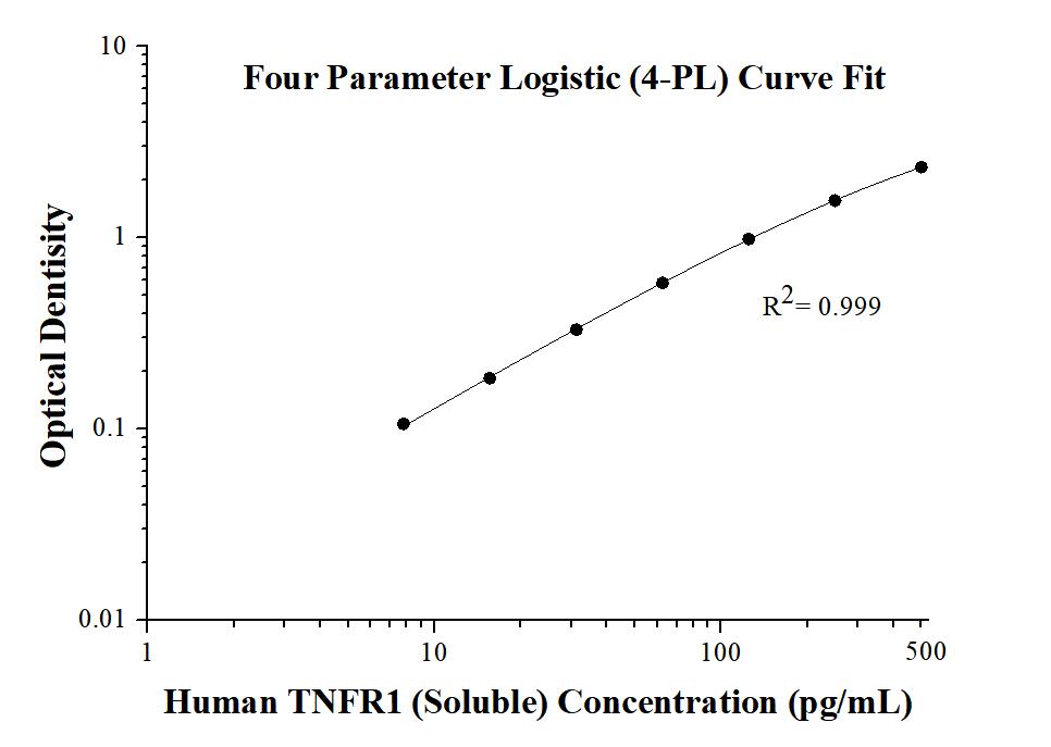
 at 0.2 μg/mL (100 μL/well) can bind Human TNF alpha (GST tag) with a linear range of 0.8-3.2 ng/mL.")