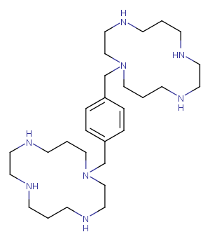
产品信息
Plerixafor, a chemokine receptor antagonist, blocks the binding of stromal cell-derived factor (SDF-1alpha) to the cellular receptor CXCR4.
|
CAS号
|
110078-46-1 |
| 分子式 |
C28H54N8 |
| 主要靶点 |
CXCR|HIV Protease|Virus Protease |
| 主要通路 |
蛋白酶体|自噬|G蛋白偶联受体|微生物学|免疫与炎症 |
| 分子量 |
502.78 |
| 纯度 |
100.00%, 此纯度可做参考,具体纯度与批次有关系,可咨询客服 |
| 储存条件 |
Powder: -20°C for 3 years | In solvent: -80°C for 1 year |
| 别名 |
普乐沙福|AMD 3100|JM3100|AMD-3329 |
靶点活性
CXCR4:44 nM|CXCL12:5.7 nM
体内活性
A single topical application of Plerixafor promotes wound healing in diabetic mice by increasing cytokine production, mobilizing bone marrow EPCs, and enhancing the activity of fibroblasts and monocytes/macrophages, thereby increasing both angiogenesis and vasculogenesis. [3] Cohorts of mice are administered with PBS, IGF1, PDGF, SCF, or VEGF for five consecutive days and Plerixafor on the 5th day. The number and size of the colonies are highest in IGF1 plus Plerixafor injected mice compared to PDGF, SCF and VEGF treated groups, in combination with Plerixafor. [4]
体外活性
Plerixafor inhibits CXCL12-mediated chemotaxis with a potency lightly better than its affinity for CXCR4. [1] Plerixafor also antagonizes SDF-1/CXCL12 ligand binding with an IC50 of 651 nM. Plerixafor inhibits SDF-1 mediated GTP-binding, SDF-1 mediated calcium flux and SDF-1 stimulated chemotaxis with IC50 of 27 nM, 572 nM and 51 nM, respectively. Plerixafor does not inhibit calcium flux against cells expressing CXCR3, CCR1, CCR2b, CCR4, CCR5 or CCR7 when stimulated with their cognate ligands, nor does Plerixafor inhibit receptor binding of LTB4. Plerixafor does not, on its own, induce a calcium flux in the CCRF–CEM cells, which express multiple GPCRs including CXCR4, CCR4 and CCR7. [2]
溶解度
Ethanol:50 mg/mL,Sonification is recommended.,DMSO:2 mM,Sonification is recommended.,PBS:1mg/mL (1.98 mM),Need ultrasound,H2O:Insoluble
细胞实验
Plerixafor is dissolved in DMSO and then diluted with appropriate medium[2]. U87 mg cells are seeded in 96-well plates at the density of 6×10³ cells in 200 μL/well and treated with CXCL12, Plerixafor or with peptide R, as described in the previous "Treatments" section. MTT (5 μg/mL) is added at each time point (24, 48, 72 h) during the final 2 h of treatment. After removing cell medium, 100 μL DMSO are added and optical densities measured at 595 nm with a LT-4000MS Microplate Reader. Measurements are made in triplicates from three independent experiments[2].

