Recombinant Mouse uPAR/CD87 protein (rFc Tag)
种属
Mouse
纯度
>90 %, SDS-PAGE
标签
rFc Tag
生物活性
未测试
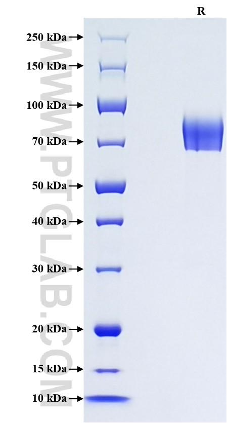
验证数据展示
产品信息
| 纯度 | >90 %, SDS-PAGE |
| 内毒素 | <0.1 EU/μg protein, LAL method |
| 生物活性 |
Not tested |
| 来源 | HEK293-derived Mouse uPAR protein Leu24-Thr297 (Accession# P35456-1) with a rabbit IgG Fc tag at the C-terminus. |
| 基因ID | 18793 |
| 蛋白编号 | P35456-1 |
| 预测分子量 | 56.0 kDa |
| SDS-PAGE | 65-90 kDa, reducing (R) conditions |
| 组分 | Lyophilized from 0.22 μm filtered solution in PBS, pH 7.4. Normally 5% trehalose and 5% mannitol are added as protectants before lyophilization. |
| 复溶 | Briefly centrifuge the tube before opening. Reconstitute at 0.1-0.5 mg/mL in sterile water. |
| 储存条件 |
It is recommended that the protein be aliquoted for optimal storage. Avoid repeated freeze-thaw cycles.
|
| 运输条件 | The product is shipped at ambient temperature. Upon receipt, store it immediately at the recommended temperature. |
背景信息
uPAR/CD87 is a highly glycosylated GPI-anchored membrane protein. In addition to the membrane-anchored form, uPAR is released from the plasma membrane by cleavage of the GPI anchor and can be found as a soluble form (suPAR). uPAR contains three homologous domains (D1-D3) of which the N-terminal one (D1) represents the uPA-binding domain. After binding to uPAR, uPA cleaves plasminogen, generating the active protease plasmin which is involved in a wide variety of physiologic and pathologic processes. In addition to regulating proteolysis, uPAR has important function in cell adhesion, migration and proliferation. Studies reveal that uPAR expression is elevated during inflammation and tissue remodelling and in many human cancers, in which it frequently indicates poor prognosis. suPAR has been detected in plasma, and increased plasma concentrations of suPAR have been found in patients with some advanced cancers.
参考文献:
1. Ploug M. et al. (1991). J Biol Chem. 266(3):1926-1933.
2. Stephens RW. et al. (1997). Clin Chem. 43(10):1868-1876.
3. Blasi F. et al. (2002). Nat Rev Mol Cell Biol. 3(12):932-943.
4. Smith HW. et al. (2010). Nat Rev Mol Cell Biol. 11(1):23-36.
