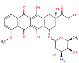
产品信息
Doxorubicin intercalation between base pairs in the DNA helix, thereby preventing DNA replication. It also inhibits topoisomerase II.
|
CAS号
|
25316-40-9 |
| 分子式 |
C27H29NO11·HCl |
| 主要靶点 |
HBV|Apoptosis|Mitophagy|Autophagy|Topoisomerase|AMPK|Antibacterial|Antibiotic|HIV Protease |
| 主要通路 |
DNA损伤和修复|蛋白酶体|表观遗传|微生物学|PI3K/Akt/mTOR信号通路|凋亡|自噬 |
| 分子量 |
579.99 |
| 纯度 |
99.13%, 此纯度可做参考,具体纯度与批次有关系,可咨询客服 |
| 储存条件 |
Powder: -20°C for 3 years | In solvent: -80°C for 1 year |
| 别名 |
盐酸阿霉素|NSC 123127|Hydroxydaunorubicin hydrochloride|盐酸多柔比星|Adriamycin|Doxorubicin (Adriamycin) HCl |
靶点活性
Topoisomerase I:0.8 μM (IC50)|Topoisomerase II:2.67 μM (IC50)
体内活性
Mice bearing PC3 xenografts are injected with 2, 4 or 8 mg/kg Doxorubicin and tumor volume is measured over time. A dose of 2 mg/kg does not affect tumor growth while higher dosages delay tumor growth initially (p<0.05 at days 18 and 22), 4 mg/kg or 8 mg/kg Doxorubicin significantly reduces levels of c-FLIP in PC3 xenografts[3]. Vehiculum, DOX or L-DOX were applied intraperitoneally in a single dose to male Wistar rats (3 groups: control, DOX, and L-DOX, respectively). Rats were sacrificed after 24 h, and samples from left atrium (LA)/ventricle (LV) and right atrium (RA)/ventricle (RV) were obtained. Expressions of miR-208a, let-7g, and snU6 were determined using qRT-PCR. In the control group, miR-208a was highly abundant in the atria compared to the ventricles and in the left-sided structures compared to the right-sided structures, while let-7g showed only atrio-ventricular gradient with predominant expression in the atria. Administration of both DOX and L-DOX resulted in 38.87 and 23.57% reduction in miR-208a expression in the LV (p = 0.028) and in 13.79 and 14.70% reduction in let-7g expression in the LA (p = 0.015), respectively. Acute administration of DOX/L-DOX alters expression of miR-208a in LV and of let-7g in LA [4].
体外活性
Doxorubicin (Dox) triggered a marked accumulation of p53 and induced CD95 gene expression and apoptosis in proliferating human umbilical vein endothelial cells (HUVECs). Transfection and site-directed mutagenesis experiments using the CD95 promoter fused to an intronic enhancer indicated the requirement for a p53 site for Dox-induced promoter activation. Furthermore, the p53 inhibitor pifithrin-alpha (PFT-alpha) blocked both promoter inducibility and protein up-regulation of CD95 in response to Dox. Up-regulated CD95 in Dox-treated cells was functional in eliciting apoptosis upon incubation of the cells with an agonistic CD95 antibody [1]. Combination of Doxorubicin and Simvastatin in the highest tested concentrations (2 μM and 10 μM, respectively) kills 97% of the Hela cells [2].
溶解度
H2O:29 mg/mL (50 mM),DMSO:29 mg/mL (50 mM)
细胞实验
To analyze the effect of Bcl-2 expression on the viability of HUVECs treated with Dox, cells were co-transfected with 200 ng of the pEGFP-spectrin expression plasmid together with 200 ng of either pCDNA3-hBcl-2 or the control pCMVβ-galactosidase expression vector (33). The pGL3 Basic vector (2.1 μg) was added as a DNA carrier in a total volume of 0.140 ml, and transfection was performed by the calcium phosphate procedure in 35-mm tissue culture dishes. After treatment, the cells were washed with PBS, fixed with 3.7% formaldehyde for 15 min, and washed for a further 10 min with 50 mM NH4Cl blocking solution in PBS. Cells were then washed with PBS, permeabilized with a 0.1% Triton X-100 for 10 min, washed again with PBS, and stained with 1 μg/ml 4′,6-diamidino-2-phenyl-indole solution for 2 min. The cells were examined under a fluorescence microscope, and GFP-positive cells were scored after counting a minimum of 1000 total cells for each condition. The efficiency of transfection in Bcl-2- and β-galactosidase-expressing cells, determined in aliquots of transfected cells just before the addition of Dox, was similar (10–12%) [1].
动物实验
Athymic male nude mice (3-4 weeks old) are used. PC3 cells (4×10⁶) are injected subcutaneously into the flanks of mice. Animals bearing tumors are randomly assigned to treatment groups (five or six mice per group) and treatment initiated when xenografts reached volumes of about 100 mm3. Tumors are measured using digital calipers and volume calculated using the formula: Volume=Width2×Length×0.52, where width represents the shorter dimension of the tumor. Treatments are administered as indicated using vehicle (PBS containing 0.1% BSA), Doxorubicin (2-8 mg/kg), Apo2L/TRAIL (500 μg/animal), or a combination of 4 mg/kg Doxorubicin followed by 500 μg Apo2L/TRAIL. Doxorubicin is administered systemically whereas Apo2L/TRAIL is given either intratumorally or systemically. All treatments are given once. Mice are monitored daily for signs of adverse effects (listlessness and scruffy appearance). Treatments seemed to be well tolerated. The mean±SEM is calculated for each data point. Differences between treatment groups are analyzed by the student t-test. Differences are considered significant when P<0.05 [3]. Altogether, 29 male Wistar rats (weight 306 ± 18.6 g) were used in the study. Animals were divided into three groups: control (group C; n = 10; 306.4 ± 17.2 g), animals treated with DOX (group DOX; n = 10; 305.0 ± 24.9 g) and animals treated with L-DOX (group L-DOX; n = 9; 306.7 ± 15.0 g). Vehiculum (aqua pro injection), DOX and L-DOX were applied to group C, DOX and L-DOX, respectively, by single intraperitoneal injection; concentration of both DOX and L-DOX was 5 mg/kg, similar to the concentrations used in human treatment protocols. All animals were sacrificed 24 h after drug application. Thoracotomy was performed, hearts were excised and samples were obtained separately from the free wall of the left atrium (LA), left ventricle (LV), right atrium (RA) and right ventricle (RV).Samples were placed into RNA later preservation solution and stored at -80 C until further analysis [4].

