Recombinant Human Podoplanin protein (rFc Tag)(HPLC verified)
种属
Human
纯度
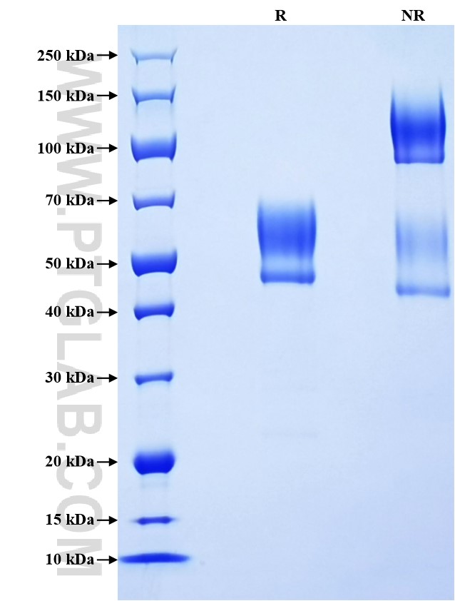
>90 %, SDS-PAGE
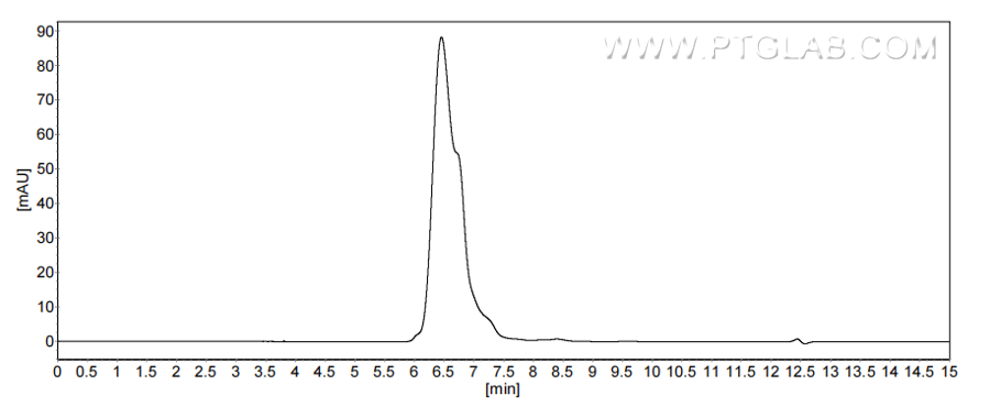
>90 %, SEC-HPLC
标签
rFc Tag
生物活性
未测试
验证数据展示
产品信息
| 纯度 | >90 %, SDS-PAGE >90 %, SEC-HPLC |
| 内毒素 | <0.1 EU/μg protein, LAL method |
| 生物活性 |
Not tested |
| 来源 | HEK293-derived Human Podoplanin protein Ala23-Leu131 (Accession# Q86YL7-1) with a rabbit IgG Fc tag at the C-terminus. |
| 基因ID | 10630 |
| 蛋白编号 | Q86YL7-1 |
| 预测分子量 | 37.4 kDa |
| SDS-PAGE | 48-68 kDa, reducing (R) conditions |
| 组分 | Lyophilized from 0.22 μm filtered solution in PBS, pH 7.4. Normally 5% trehalose and 5% mannitol are added as protectants before lyophilization. |
| 复溶 | Briefly centrifuge the tube before opening. Reconstitute at 0.1-0.5 mg/mL in sterile water. |
| 储存条件 |
It is recommended that the protein be aliquoted for optimal storage. Avoid repeated freeze-thaw cycles.
|
| 运输条件 | The product is shipped at ambient temperature. Upon receipt, store it immediately at the recommended temperature. |
背景信息
Podoplanin (PDPN) is a transmembrane glycoprotein that plays a role in a variety of biological processes including regulation of blood-lymphatic vessel development, cerebral vascular pattern and integrity, cell motility, tumorigenesis and metastasis, and mediates tumor cell-induced platelet aggregation in different cancer types. It is a lymphatic marker because the expression of podoplanin has been detected in lymphatic but not blood vascular endothelium, and is useful as the marker of tumor-associated Lymphangiogenesis. When expressed in keratinocytes, PDPN induces changes in cell morphology with transfected cells showing an elongated shape, numerous membrane protrusions, major reorganization of the actin cytoskeleton, increased motility and decreased cell adhesion.
参考文献:
1. Ukaji T, et al . Cancer Sci. 2021; 112(6):2299-2313.
2. Krishnan H, et al. J Biol Chem. 2013 ;288(17):12215-21.
3. Wang X, et al. Cancer Gene Ther. 2023 ;30(2):345-357.
4.Astarita JL, et al. Front Immunol. 2012; 3:283.


