Recombinant Human CSPG4 protein (Myc Tag, His Tag)
种属
Human
纯度
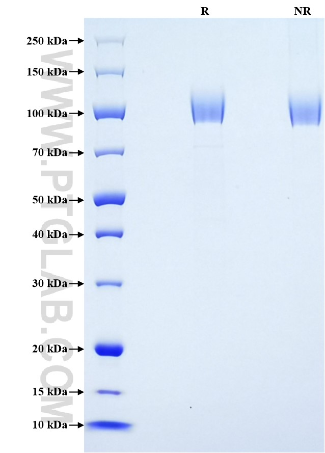
>90 %, SDS-PAGE
标签
Myc Tag, His Tag
生物活性
未测试
验证数据展示
产品信息
| 纯度 | >90 %, SDS-PAGE |
| 内毒素 | <0.1 EU/μg protein, LAL method |
| 生物活性 |
Not tested |
| 来源 | HEK293-derived Human CSPG4 protein Ser1583-Ser2224 (Accession# Q6UVK1) with a Myc tag and a His tag at the C-terminus. |
| 基因ID | 1464 |
| 蛋白编号 | Q6UVK1 |
| 预测分子量 | 73.2 kDa |
| SDS-PAGE | 90-125 kDa, reducing (R) conditions |
| 组分 | Lyophilized from 0.22 μm filtered solution in PBS, pH 7.4. Normally 5% trehalose and 5% mannitol are added as protectants before lyophilization. |
| 复溶 | Briefly centrifuge the tube before opening. Reconstitute at 0.1-0.5 mg/mL in sterile water. |
| 储存条件 |
It is recommended that the protein be aliquoted for optimal storage. Avoid repeated freeze-thaw cycles.
|
| 运输条件 | The product is shipped at ambient temperature. Upon receipt, store it immediately at the recommended temperature. |
背景信息
CSPG4, also named as HMW-MAA, MCSP, MCSPG, MEL-CSPG, MSK16 and NG2, is a proteoglycan playing a role in cell proliferation and migration which stimulates endothelial cells motility during microvascular morphogenesis. CSPG4 may inhibit neurite outgrowth and growth cone collapse during axon regeneration. It is cell surface receptor for collagen alpha 2(VI) which may confer cells ability to migrate on that substrate. CSPG4 is also a signal-transducing protein by binding through its cytoplasmic C-terminus scaffolding and signaling proteins. It is a target of specific diagnosis and immunotherapy with antiidiotypic antibodies due, in part, to its roles in growth control, adhesion, cell-substratum interactions, and cell-cell contacts.
参考文献:
1. Matthew A Price, et al. (2011) Pigment Cell Melanoma Res. 24(6):1148-57.
2. J Iida, et al. (2001) J Biol Chem. 276(22):18786-94.
3. Jianbo Yang, et al. (2004) J Cell Biol. 165(6):881-91.
4. K M Eisenmann, et al. (1999) Nat Cell Biol. 1(8):507-13.
5. Valeria Rolih, et al. (2017) J Transl Med. 15(1):151.
