- Featured Product
- KD/KO Validated
Calbindin-D28k Polyclonal antibody
Calbindin-D28k Polyclonal Antibody for IF/ICC,IF-Fro,IF-P, IHC, IP, WB, ELISA
Host / Isotype
Rabbit / IgG
Reactivity
human, mouse, rat
Applications
IF/ICC,IF-Fro,IF-P, IHC, IP, WB, ELISA
Conjugate
Unconjugated
验证数据展示
 at dilution of 1:4000 incubated at room temperature for 1.5 hours.")
 fixed mouse cerebellum tissue using Calbindin-D28k antibody (14479-1-AP) at dilution of 1:200 and CoraLite®488-Conjugated AffiniPure Goat Anti-Rabbit IgG(H+L), PAX6 antibody (<a class='green' href='/productredirect?CatalogNo=67529-1-Ig' target='_blank'>67529-1-Ig</a>, Clone: 2C5A1, red).")
 fixed mouse cerebellum tissue using Calbindin-D28k antibody (14479-1-AP) at dilution of 1:200 and CoraLite®488-Conjugated AffiniPure Goat Anti-Rabbit IgG(H+L), PAX6 antibody (<a class='green' href='/productredirect?CatalogNo=67529-1-Ig' target='_blank'>67529-1-Ig</a>, Clone: 2C5A1, red).")
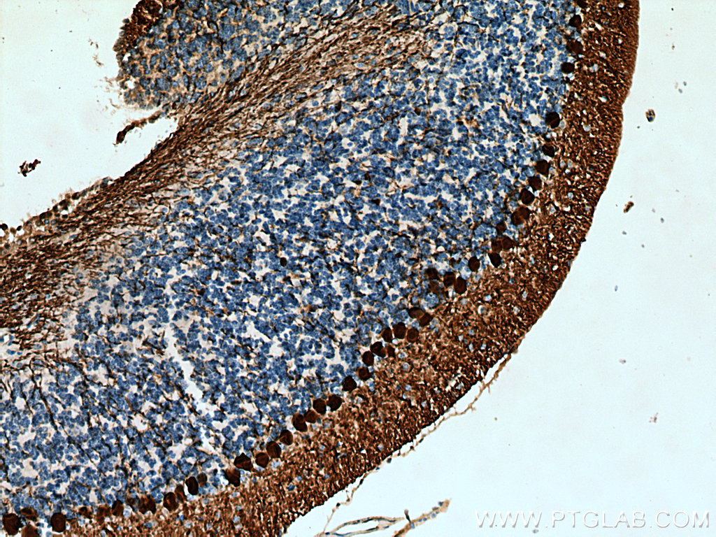
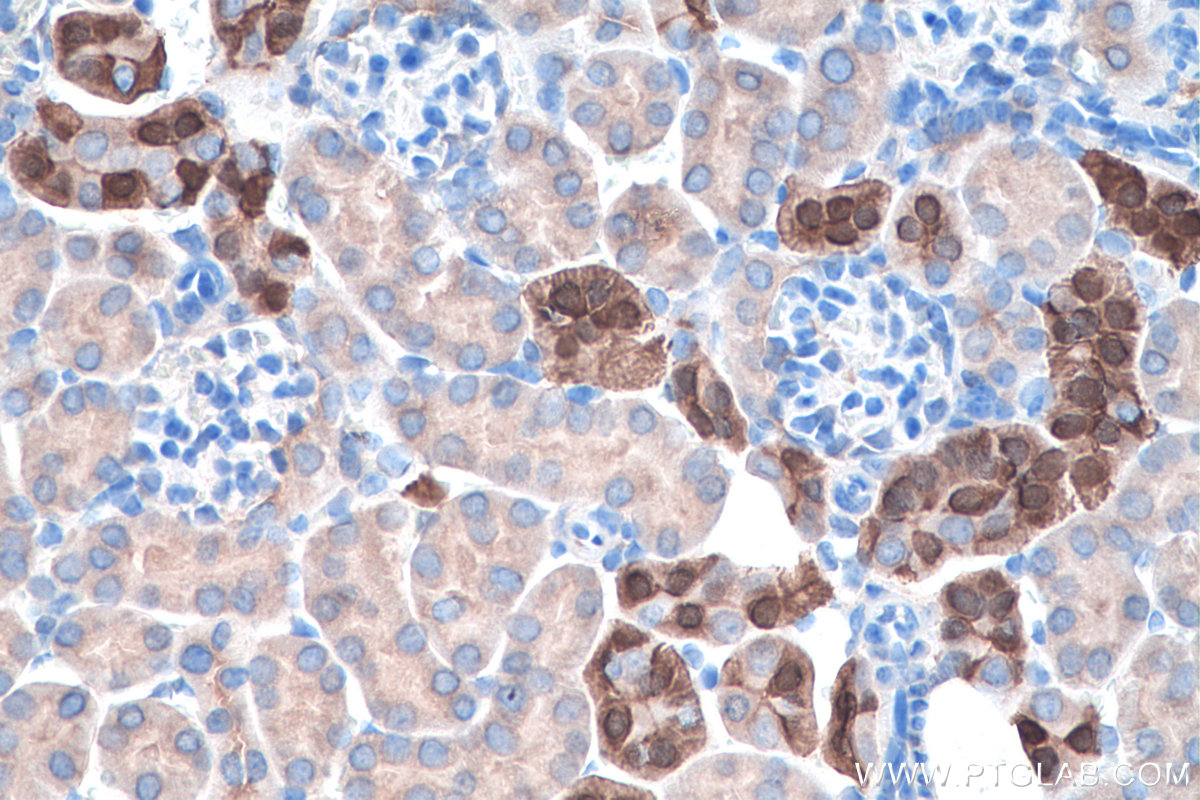
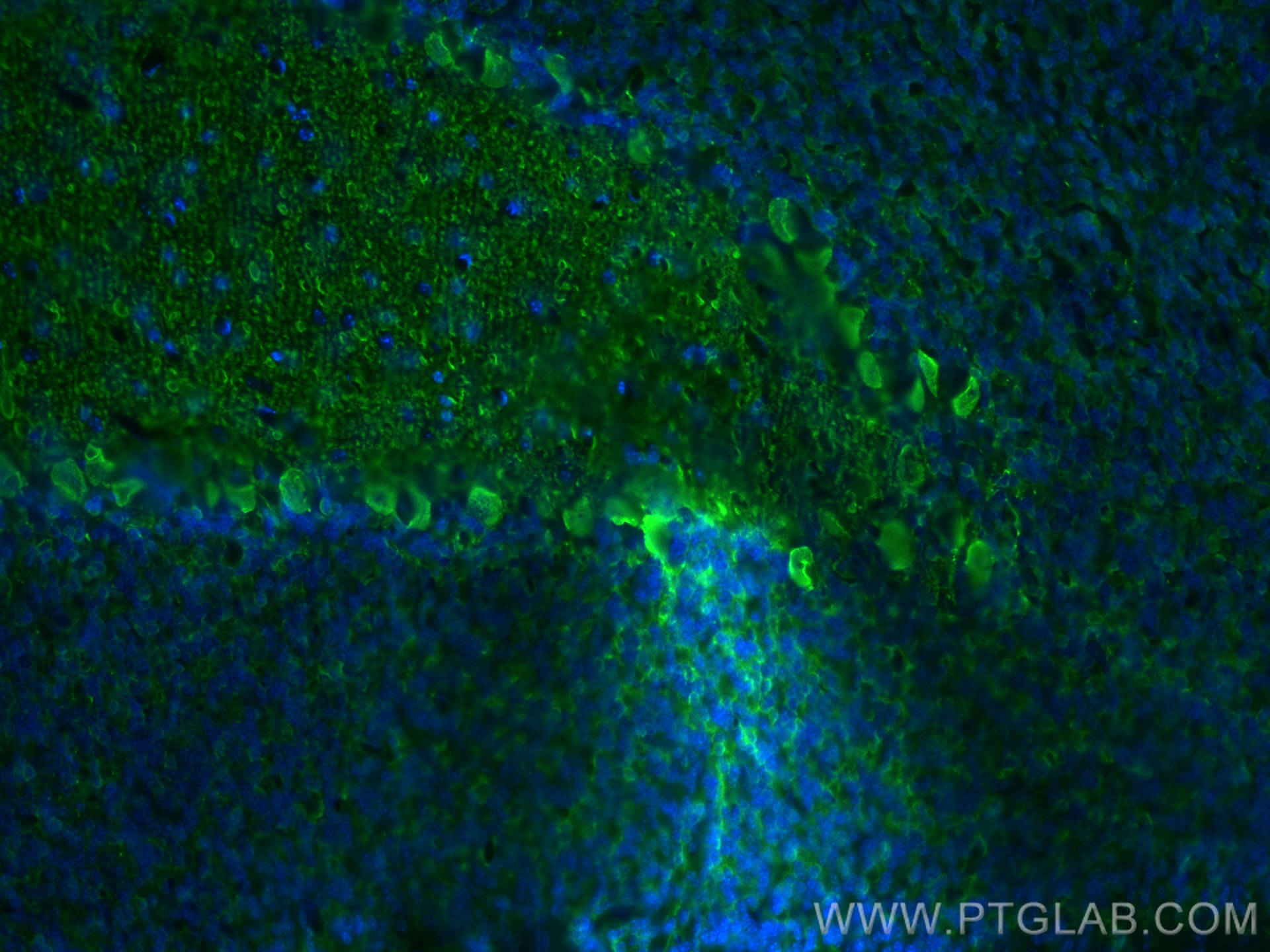
 at dilution of 1:2000 (under 10x lens).")
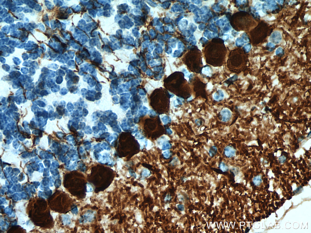
 at dilution of 1:2000 (under 40x lens).")
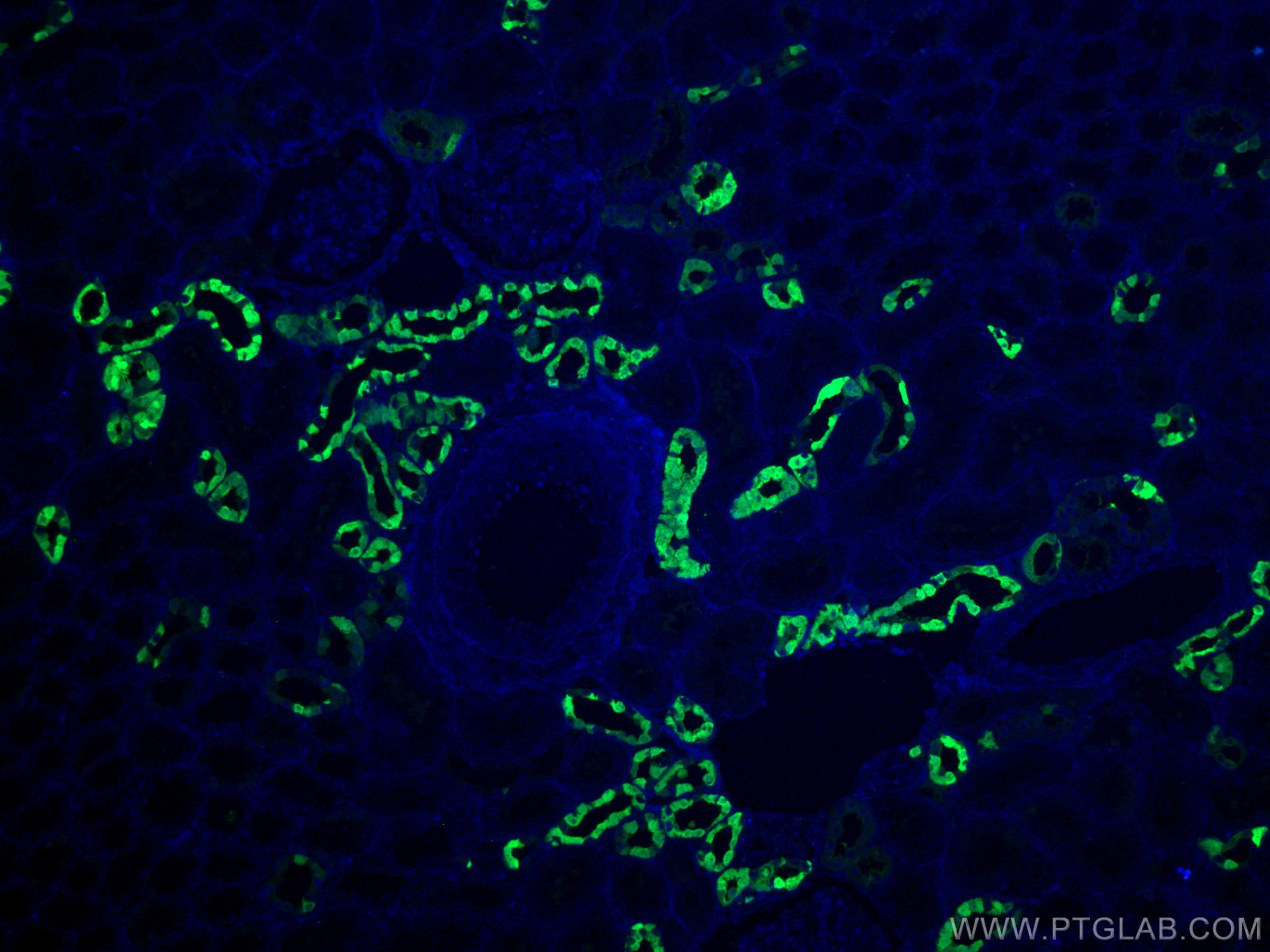
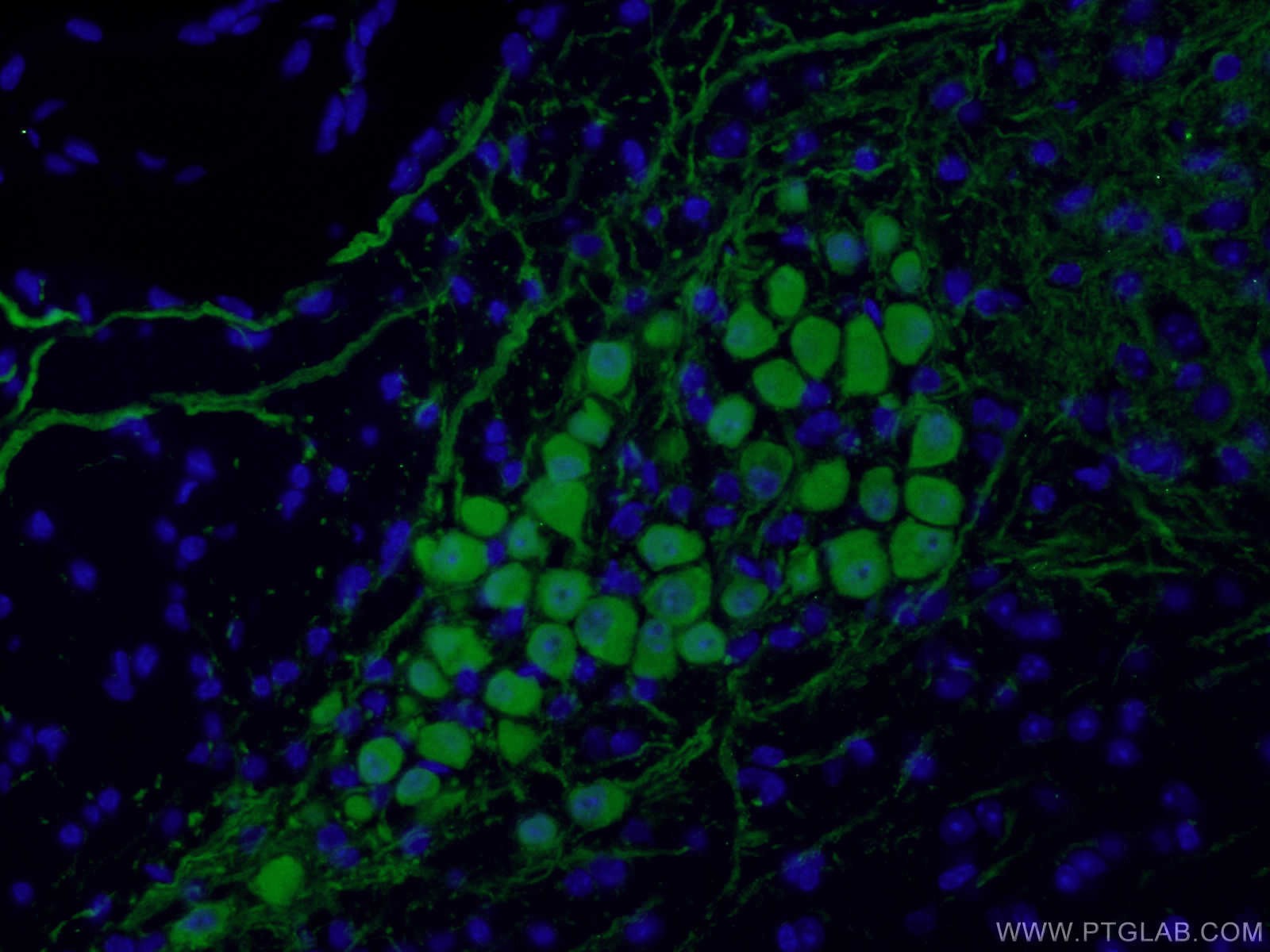
 fixed mouse cerebellum tissue using 14479-1-AP (Calbindin-D28k antibody) at dilution of 1:100 and Alexa Fluor 488-conjugated AffiniPure Goat Anti-Rabbit IgG(H+L).")
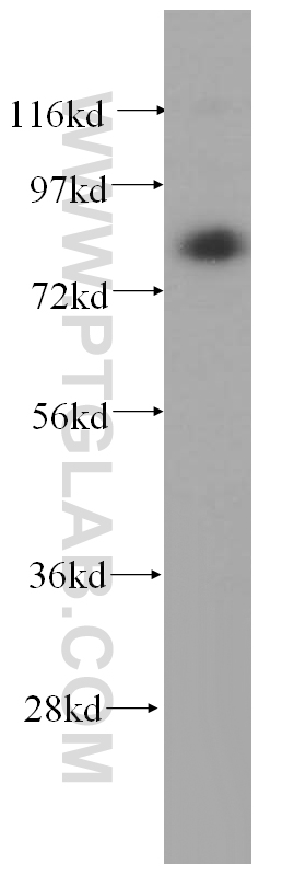
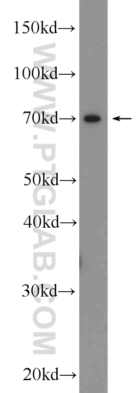
 with mouse brain tissue lysate 4000ug.")
 fixed mouse cerebellum tissue using 14479-1-AP (Calbindin-D28k antibody) at dilution of 1:100 and Alexa Fluor 488-conjugated AffiniPure Goat Anti-Rabbit IgG(H+L).")
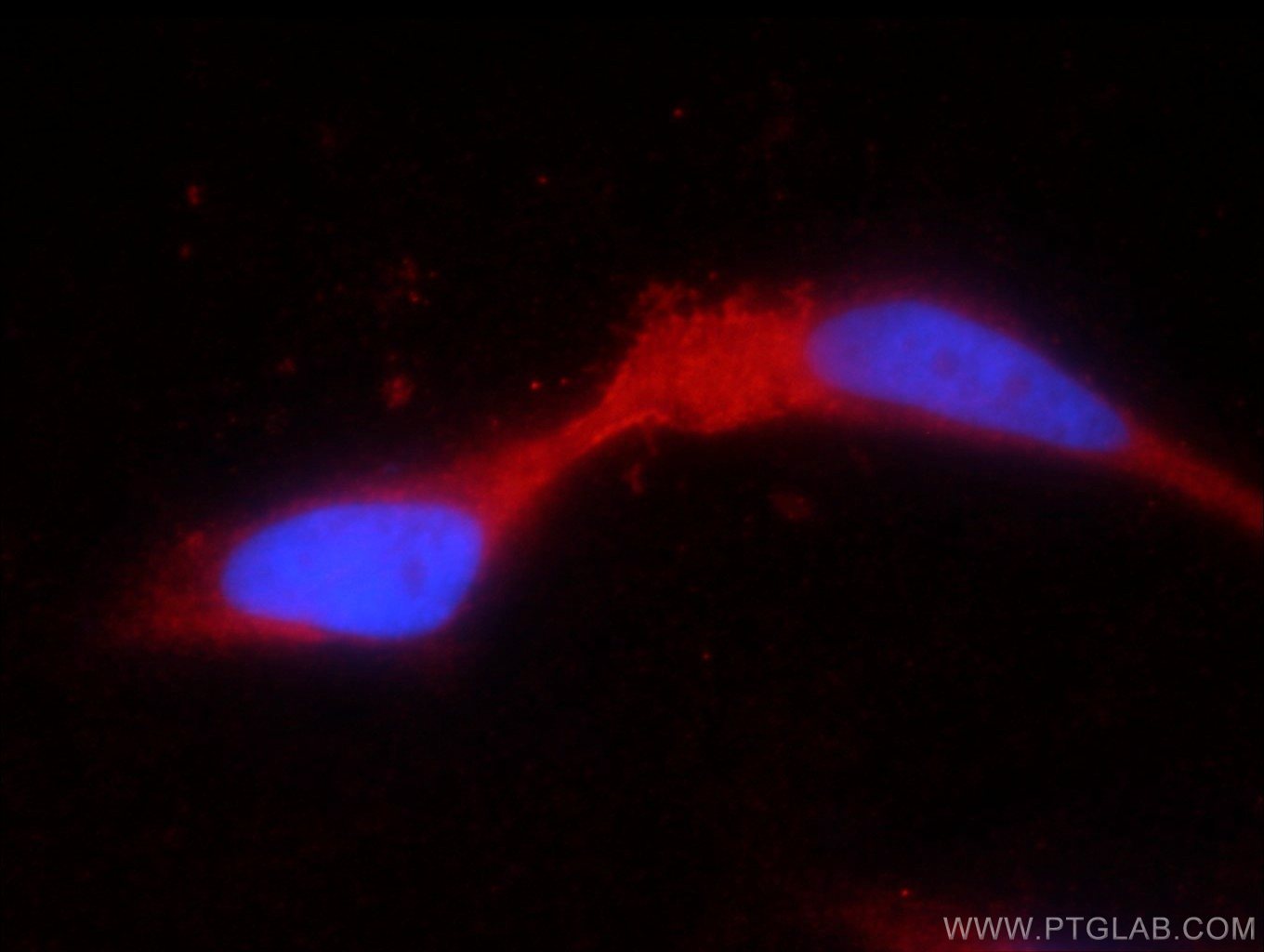
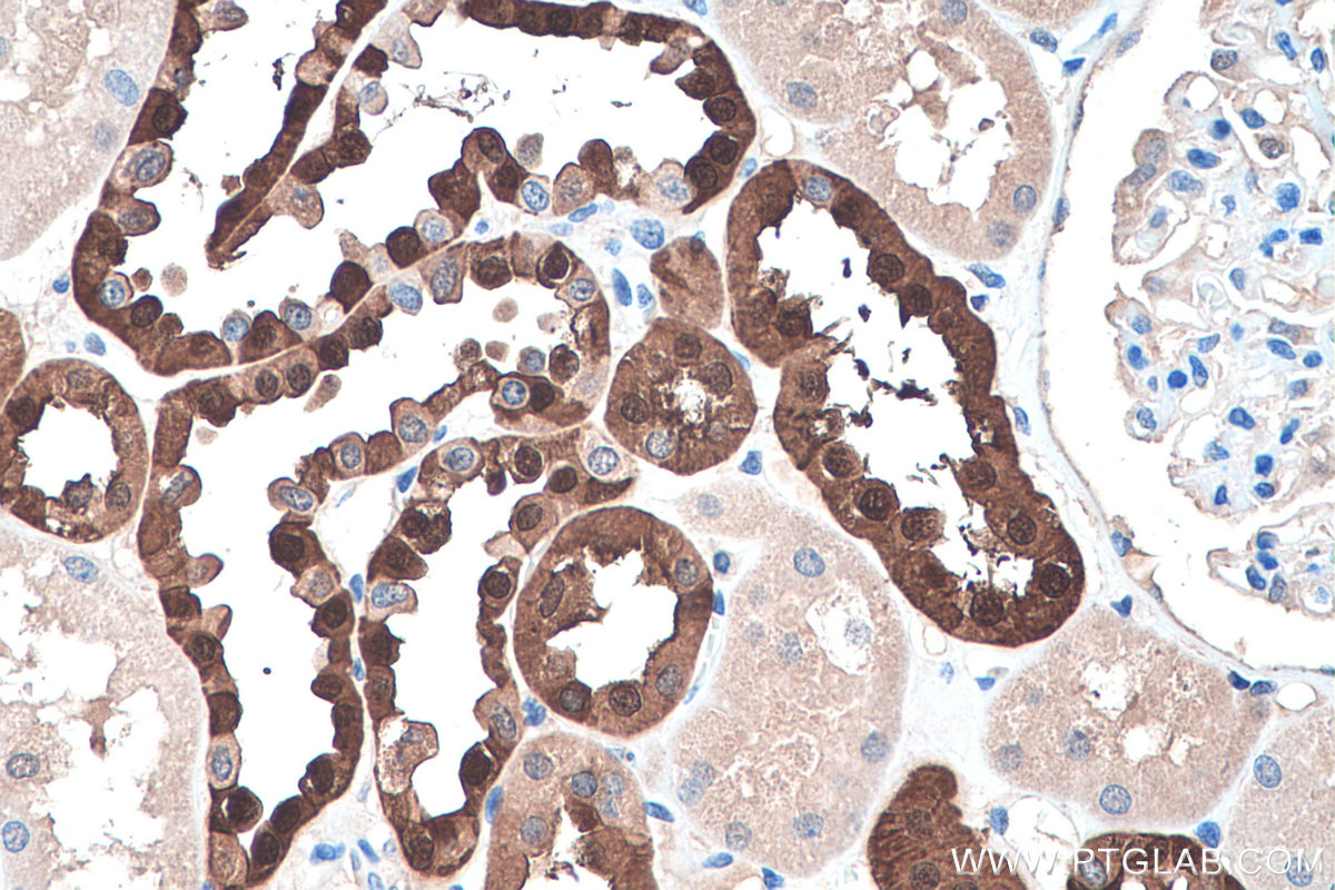
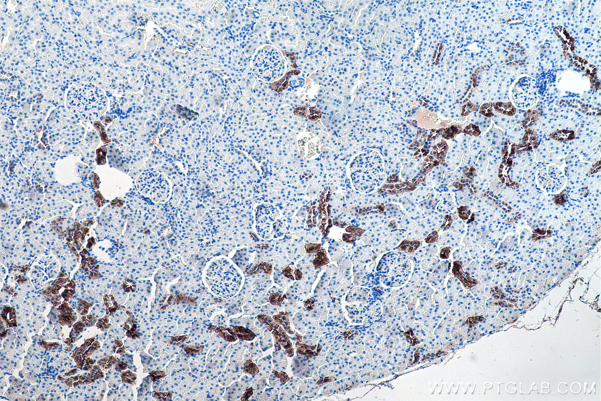
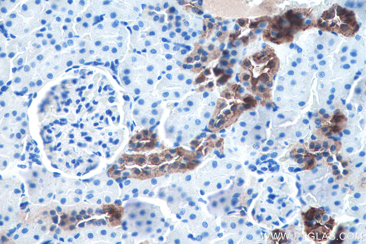
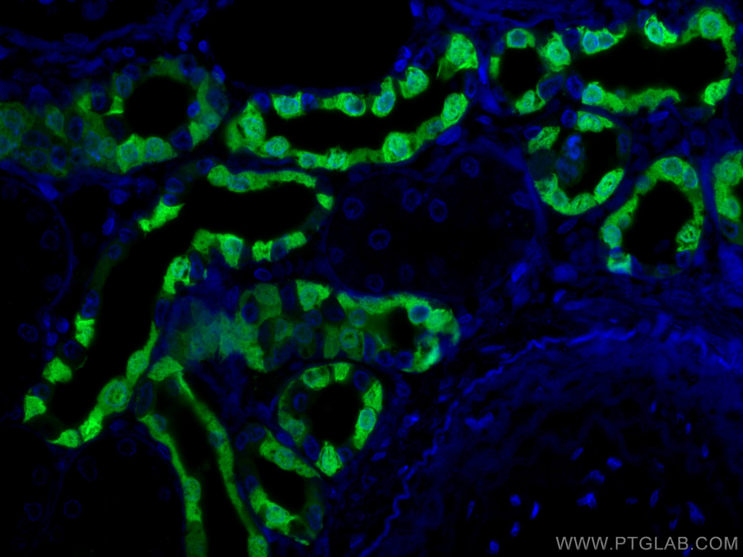
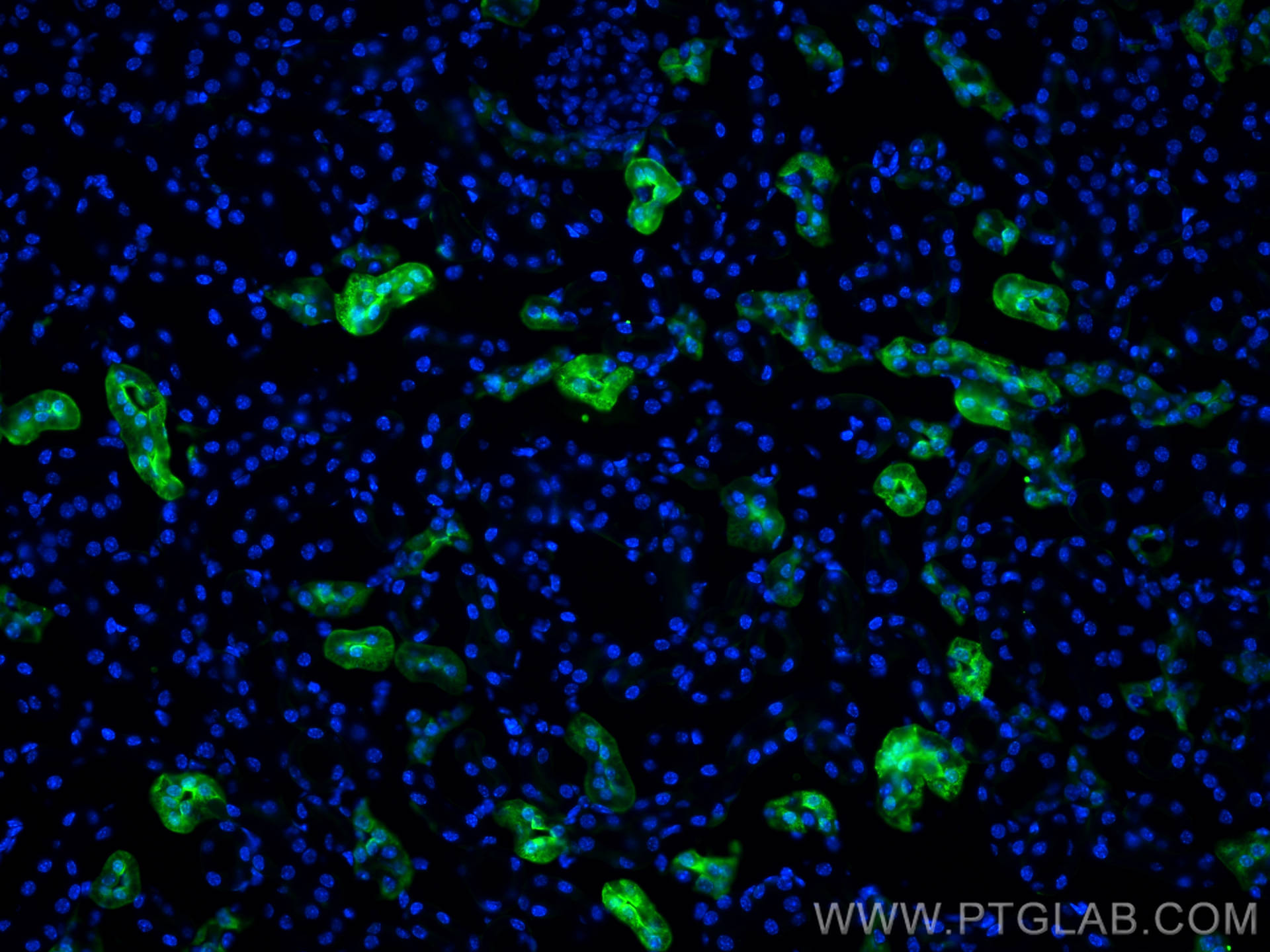
 fixed human kidney tissue using Calbindin-D28k antibody (14479-1-AP) at dilution of 1:200 and CoraLite®488-Conjugated AffiniPure Goat Anti-Rabbit IgG(H+L), PTPRO antibody (<a class='green' href='/productredirect?CatalogNo=67000-1-Ig' target='_blank'>67000-1-Ig</a>, Clone: 2F2B4, red).")
经过测试的应用
| Positive WB detected in | mouse brain tissue, human kidney tissue, human brain tissue, rat brain tissue |
| Positive IP detected in | mouse brain tissue |
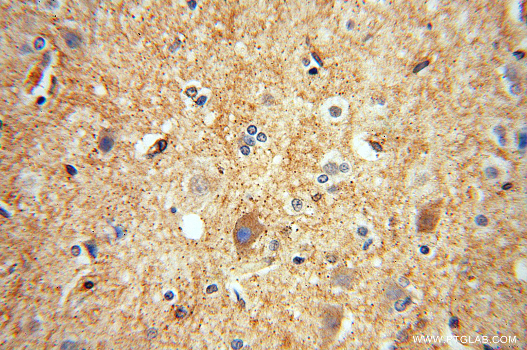
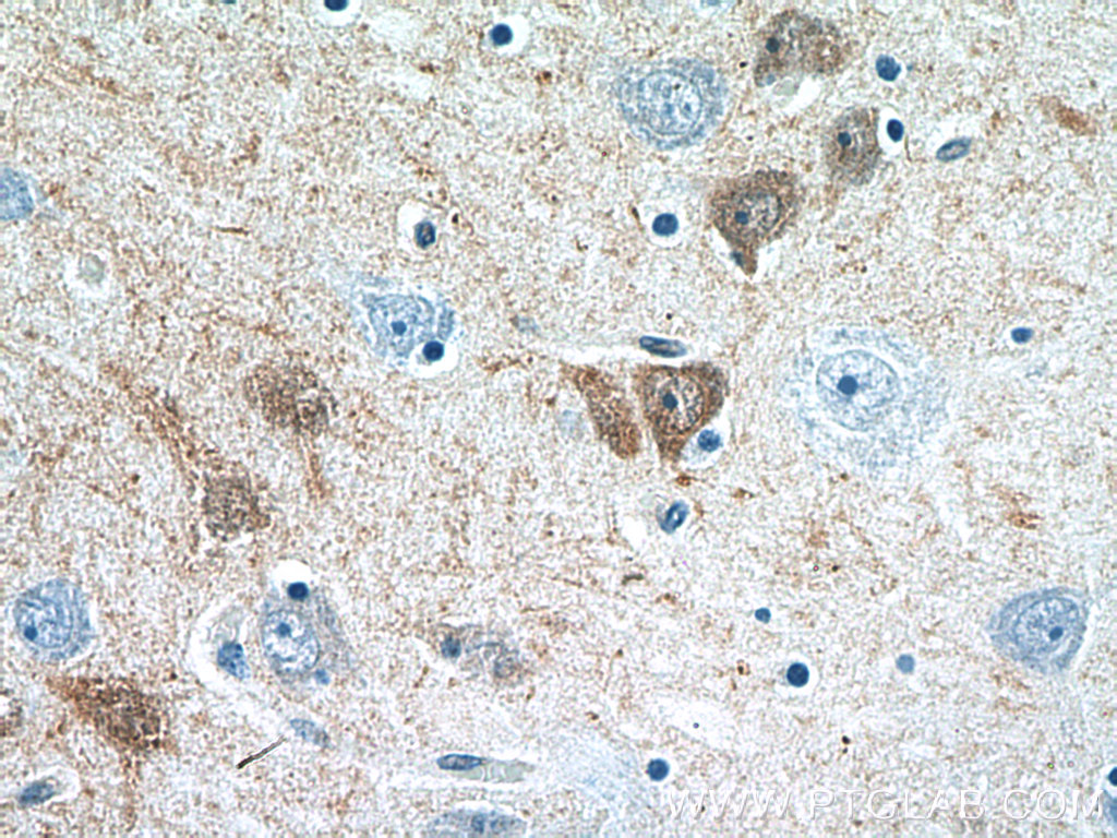
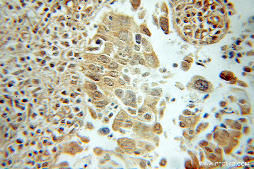
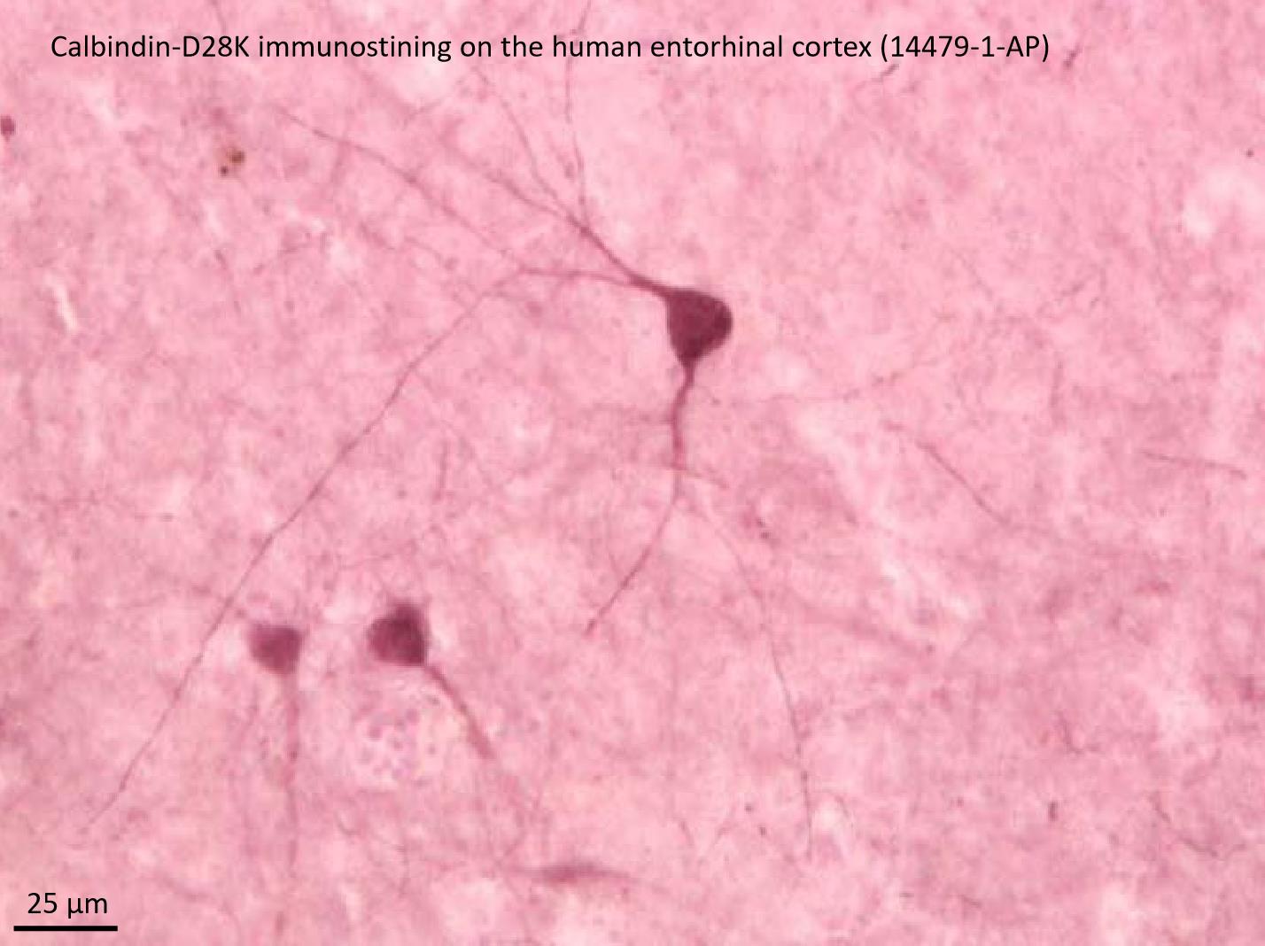
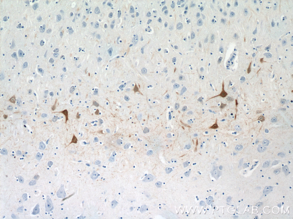
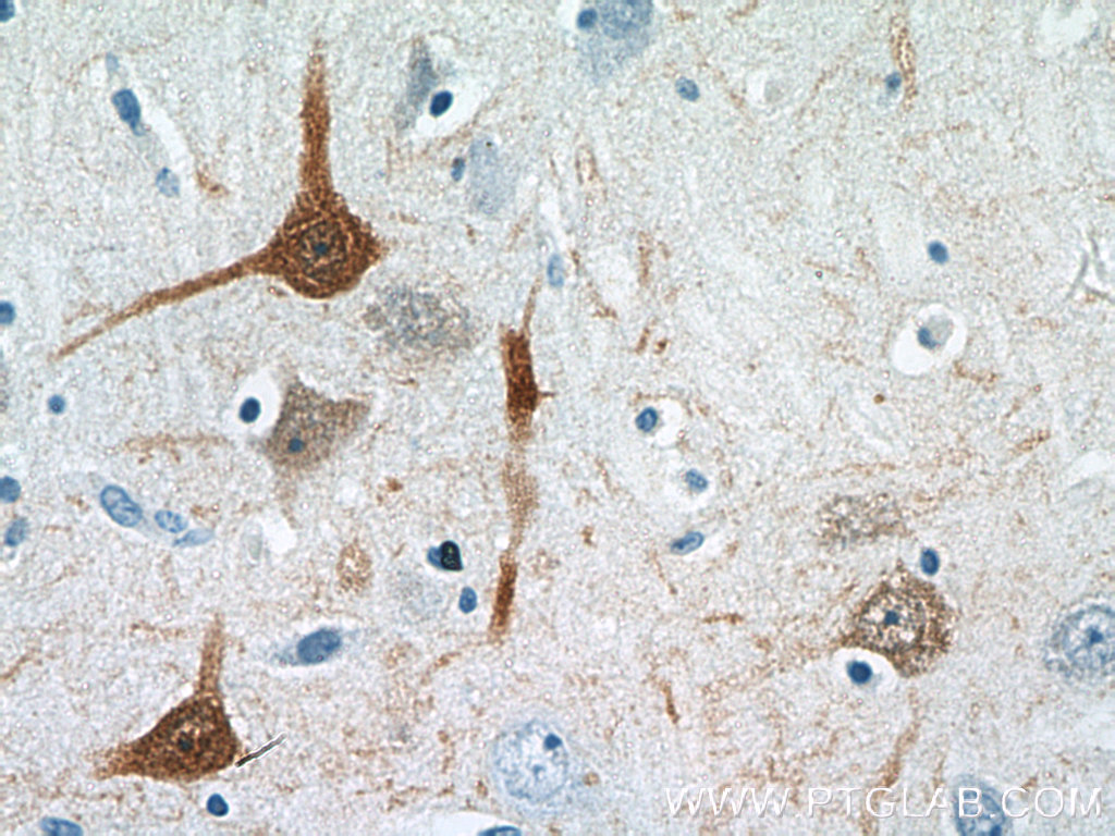
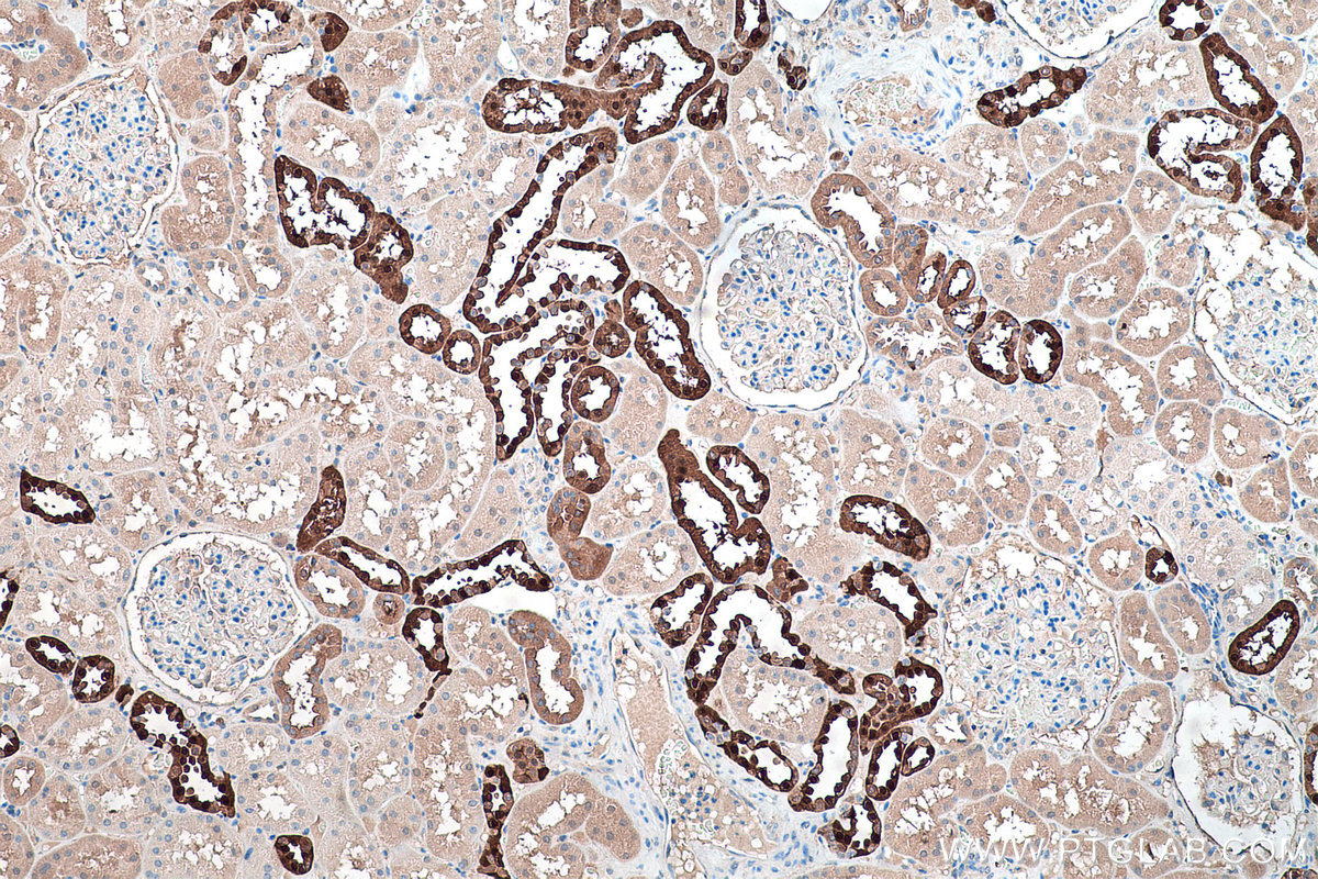
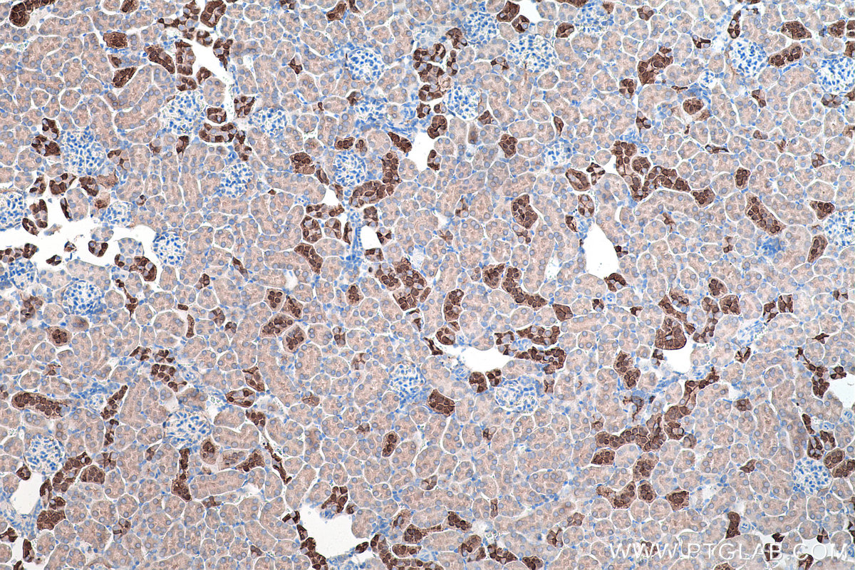
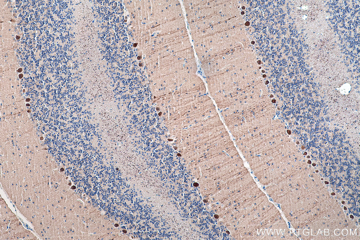
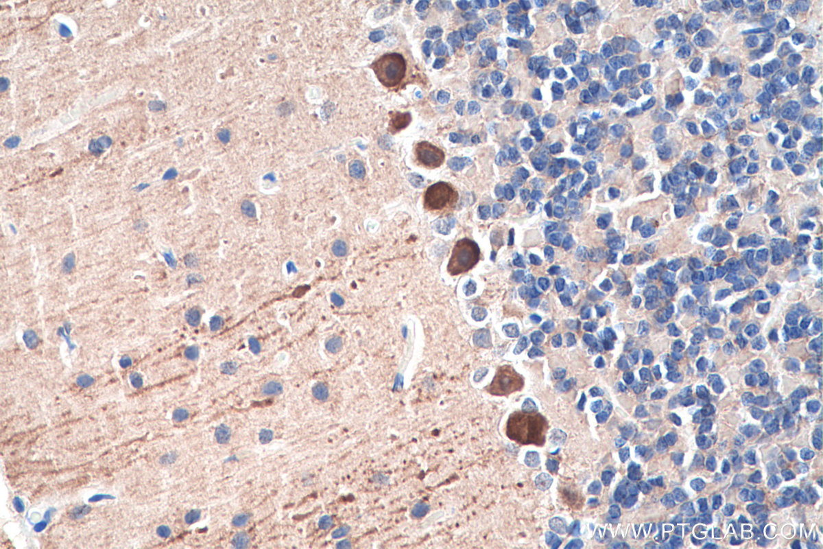
| Positive IHC detected in | human cerebellum tissue, hippocampus, human brain tissue, mouse cerebellum tissue, rat cerebellum tissue, human kidney tissue, mouse kidney tissue, rat kidney tissue Note: suggested antigen retrieval with TE buffer pH 9.0; (*) Alternatively, antigen retrieval may be performed with citrate buffer pH 6.0 |
| Positive IF detected in | mouse cerebellum tissue, human kidney tissue, mouse kidney tissue |
Planning an IHC experiment? We recommend our IHCeasy Calbindin-D28k Ready-To-Use IHC Kit. Calbindin-D28k primary antibody included.
Planning an IF experiment? We recommend our CoraLite® Plus 488 and CoraLite®594 conjugated versions of this antibody.
推荐稀释比
| Application | Dilution |
|---|---|
| Western Blot (WB) | WB : 1:1000-1:8000 |
| Immunoprecipitation (IP) | IP : 0.5-4.0 ug for 1.0-3.0 mg of total protein lysate |
| Immunohistochemistry (IHC) | IHC : 1:1000-1:4000 |
| Immunofluorescence (IF) | IF : 1:50-1:500 |
| It is recommended that this reagent should be titrated in each testing system to obtain optimal results. | |
| Sample-dependent, Check data in validation data gallery. | |
产品信息
14479-1-AP targets Calbindin-D28k in WB, IP, IF, IHC, ELISA applications and shows reactivity with human, mouse, rat samples.
| Tested Applications | IF/ICC,IF-Fro,IF-P, IHC, IP, WB, ELISA |
| Cited Applications | WB, IF, IHC |
| Tested Reactivity | human, mouse, rat |
| Cited Reactivity | human, mouse, rat |
| Immunogen | Calbindin-D28k fusion protein Ag5861 种属同源性预测 |
| Host / Isotype | Rabbit / IgG |
| Class | Polyclonal |
| Type | Antibody |
| Full Name | calbindin 1, 28kDa |
| Synonyms | CAB27, CALB, CALB1, Calbindin, calbindin 1, 28kDa, Calbindin D28, Calbindin-D28k, D 28K |
| Calculated Molecular Weight | 30 kDa |
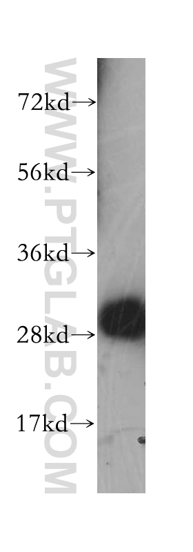
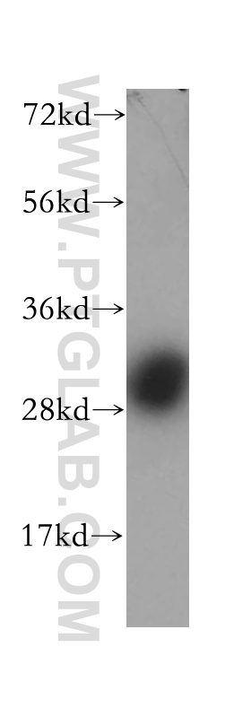
| Observed Molecular Weight | 28 kDa |
| GenBank Accession Number | BC006478 |
| Gene Symbol | CALB1 |
| Gene ID (NCBI) | 793 |
| RRID | AB_2228318 |
| Conjugate | Unconjugated |
| Form | Liquid |
| Purification Method | Antigen affinity purification |
| UNIPROT ID | P05937 |
| Storage Buffer | PBS with 0.02% sodium azide and 50% glycerol pH 7.3. |
| Storage Conditions | Store at -20°C. Stable for one year after shipment. Aliquoting is unnecessary for -20oC storage. |
背景介绍
Calbindin-D28k is a member of the calcium-binding protein superfamily that includes calmodulin and troponin C. It is a cytosolic calcium binding protein highly expressed in the distal tubule, intestines, central nervous system, primary murine osteoblast cells and in several other organs. It plays an important role in the intracellular calcium homeostasis, its strong buffering capacity prevents cytotoxic effect of high concentration of free calcium in kidney, brain, pancreas, and intestine.
实验方案
| Product Specific Protocols | |
|---|---|
| WB protocol for Calbindin-D28k antibody 14479-1-AP | Download protocol |
| IHC protocol for Calbindin-D28k antibody 14479-1-AP | Download protocol |
| IF protocol for Calbindin-D28k antibody 14479-1-AP | Download protocol |
| IP protocol for Calbindin-D28k antibody 14479-1-AP | Download protocol |
| Standard Protocols | |
|---|---|
| Click here to view our Standard Protocols |
发表文章
| Species | Application | Title |
|---|---|---|
Cell Glia-to-Neuron Conversion by CRISPR-CasRx Alleviates Symptoms of Neurological Disease in Mice. | ||
Nat Neurosci Aerobic glycolysis is the predominant means of glucose metabolism in neuronal somata, which protects against oxidative damage | ||
Cell Death Differ The long noncoding RNA Synage regulates synapse stability and neuronal function in the cerebellum. | ||
Mol Ther sEVsRVG selectively delivers antiviral siRNA to fetus brain, inhibits ZIKV infection and mitigates ZIKV-induced microcephaly in mouse model. |